Peer Review Status: Expert-reviewed | Last Updated: April 2026
Target Audience: Radiologists, MRI Physicists, Radiology Administrators, Neuroradiologists
🔑 Key Takeaways
- Deep learning-based image reconstruction (DLBIR) can reduce MRI scan times by 21–85% while maintaining or improving image quality, depending on sequence and anatomy.
- All major MRI vendors now offer commercial DLBIR products: Siemens Deep Resolve, GE AIR Recon DL, Philips SmartSpeed AI, Canon AiCE.
- A multicenter Lancet Oncology study demonstrated that 10× acceleration preserved image quality and biomarker accuracy for glioblastoma treatment response assessment.
- Clinical impact: improved SNR, CNR, lesion conspicuity, reduced motion artifacts, and potential for sedation-free pediatric MRI.
- Key concern: hallucination risk — DL models can invent or remove lesions. Radiologists must maintain awareness of this limitation.
Background
Magnetic resonance imaging provides unparalleled soft-tissue contrast and is indispensable for neuroimaging, musculoskeletal assessment, abdominal evaluation, and cardiac imaging. However, MRI’s clinical utility is fundamentally constrained by long acquisition times — a typical brain MRI protocol takes 20–45 minutes, an abdominal MRI 30–60 minutes, and a cardiac MRI up to 90 minutes. These extended scan times increase patient discomfort and motion artifacts, limit throughput and access, drive up costs, and in pediatric and elderly populations often necessitate sedation or general anesthesia [1].
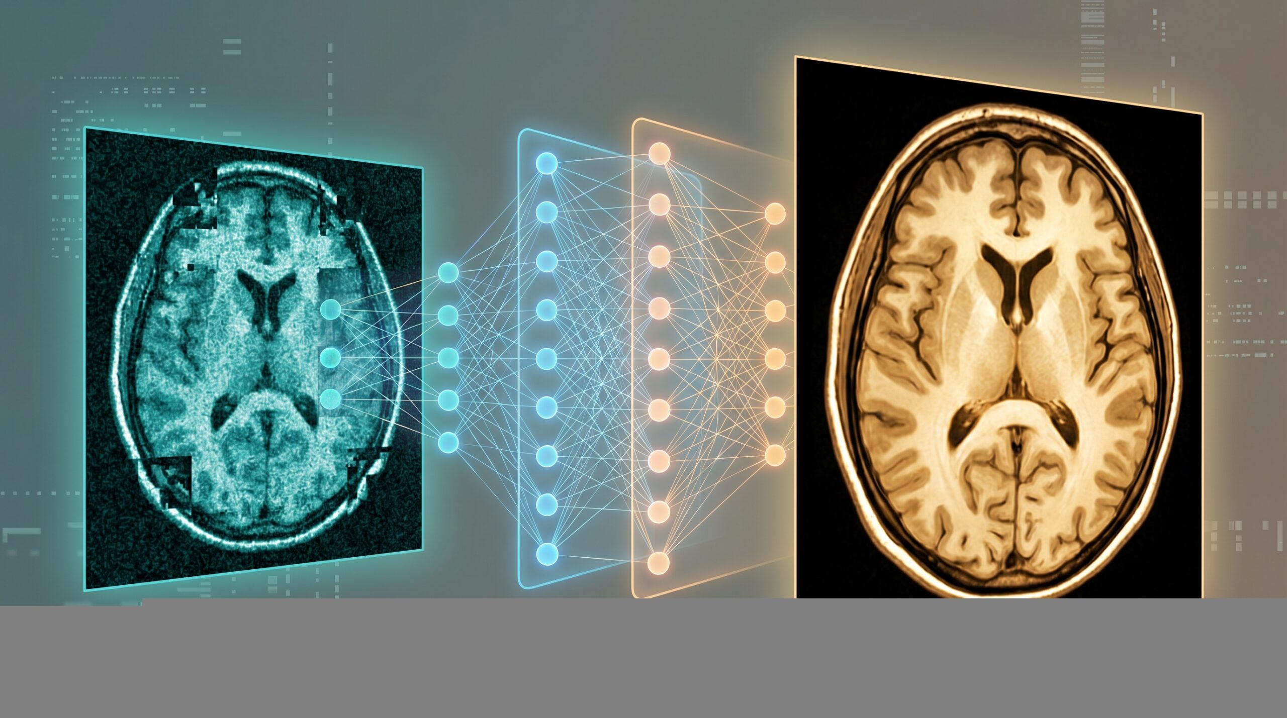
Traditional acceleration strategies — parallel imaging (GRAPPA, SENSE) and compressed sensing — reduce scan times by undersampling k-space data, but at the cost of reduced signal-to-noise ratio (SNR) and potential artifact introduction. Deep learning-based image reconstruction (DLBIR) represents a paradigm shift: neural networks trained on large datasets learn to reconstruct high-quality images from substantially undersampled data, achieving acceleration factors of 4–10× while preserving or even improving diagnostic image quality [2, 3]. This review examines the current state of DLBIR technology, vendor implementations, clinical evidence, and practical considerations for radiology departments in 2026.
How Deep Learning MRI Reconstruction Works
DLBIR approaches can be broadly categorized by where in the reconstruction pipeline the deep learning operates:
- K-space domain: Neural networks operate directly on raw acquired data, learning to fill in missing k-space samples from undersampled acquisitions. These tools are typically integrated into the MRI scanner’s reconstruction pipeline and may require newer hardware/software. Examples include physics-informed unrolled networks that alternate between data consistency and learned denoising steps [2].
- Image domain: Neural networks post-process reconstructed images to reduce noise, sharpen edges, and suppress artifacts. These tools can be applied to any MRI scanner’s output and are generally vendor-agnostic. The unenhanced images can often be reviewed alongside enhanced versions, increasing reader confidence [4].
- Hybrid approaches: Combine k-space and image-domain processing for optimal results. Most commercial vendor implementations use hybrid architectures [3].
Vendor Implementations
Table 1. Commercial Deep Learning MRI Reconstruction Products (2026)
| Vendor | Product | Approach | Claimed Time Reduction | FDA Status |
|---|---|---|---|---|
| Siemens Healthineers | Deep Resolve (Gain, Sharp, Swift, Boost) | Image + k-space hybrid; raw data-based | Up to 85% | Cleared |
| GE HealthCare | AIR Recon DL | Deep CNN in raw data space | Up to 50% | Cleared |
| Philips | SmartSpeed AI | AI-assisted compressed sensing | Up to 75% | Cleared |
| Canon Medical | AiCE / Precise IQ Engine | Deep learning denoising + super-resolution | Up to 60% | Cleared |
CNN = convolutional neural network. All products are FDA-cleared for clinical use. Actual time reductions depend on sequence, anatomy, field strength, and clinical protocol. Sources: [3, 5, 6].
Clinical Evidence
Neuroradiology
A comprehensive review in the American Journal of Neuroradiology (AJNR, January 2026) documented the current landscape of DLBIR in neuroradiology, finding that commercial implementations can achieve gradient time reductions of up to 85% while maintaining or enhancing lesion conspicuity, with improved noise suppression and diagnostic accuracy [5]. Specific applications include accelerated brain MRI without sedation in infants (using zero-echo-time silent sequences enhanced by DLBIR), improved detection of brain microbleeds on T2*-weighted imaging via super-resolution reconstruction, and faster high-resolution 3D MR neurography [5].
Body and Abdominal MRI
A 2025 review by Rajamohan et al. showed that DLBIR acceleration achieved scan time reductions of 21–93% for T2-weighted imaging and 24–62% for diffusion-weighted imaging in abdominal applications compared with conventional approaches — with improved SNR, contrast-to-noise ratio (CNR), and lesion conspicuity across studies [4]. Single-breath-hold abdominal HASTE sequences with DLBIR produced image quality equivalent to or better than conventional multi-breath-hold acquisitions at 3T [7].
Oncology: The Landmark Multicenter Study
A multicenter, retrospective cohort study published in The Lancet Oncology demonstrated that deep learning-based reconstruction of undersampled brain MRI data from over 2,000 glioblastoma patients across 200+ institutions achieved excellent image quality and preserved the accuracy of derived imaging biomarkers even at 10× acceleration — meaning a 30-minute brain MRI could theoretically be reduced to 3 minutes without compromising treatment response assessment [8]. The study’s open-source approach provides a blueprint for building similar models across body regions.
Figure 1. Clinical Impact of Deep Learning MRI Reconstruction
Scan Time
−50–85%
Depending on sequence
and anatomy
Image Quality
▲ SNR/CNR
Improved or maintained
vs. conventional
Pediatric
No sedation
Fast enough to avoid
GA in many children
Throughput
+30–50%
More patients/day
same scanner
Sources: Haller et al., Radiology 2025 [3]; Rai et al., AJNR 2026 [5]; Rajamohan et al., JCAT 2025 [4].
Challenges and Limitations
- Hallucination risk: The most clinically concerning limitation. DL models can invent lesions that do not exist (false positives) or remove true lesions from reconstructed images (false negatives). While some hallucinated artifacts are easily recognized by experienced radiologists as artifactual, subtle hallucinations in the size or morphology of real lesions could affect diagnostic accuracy. Ongoing research focuses on uncertainty quantification and detection of reconstruction artifacts [4, 9].
- Vendor specificity: Most commercial DLBIR products are vendor-specific and may require newer scanner hardware or software platforms, limiting availability on older equipment. Vendor-neutral platforms are in development but not yet widely available [5].
- Limited clinical validation at extreme acceleration: While acceleration factors of 2–4× are well-validated, factors of 8–15× remain primarily research-grade. The clinical safety and diagnostic equivalence of extreme acceleration require prospective validation in larger, diverse patient populations [8].
- Training data limitations: Models trained predominantly on data from specific scanners, field strengths, or patient populations may underperform in different clinical settings. Generalizability across institutions, protocols, and pathologies is a persistent challenge [2].
- Quantitative imaging impact: DLBIR may alter quantitative MRI biomarkers (T1/T2 mapping, ADC values, perfusion parameters). Validation of quantitative accuracy — not just visual image quality — is essential before using DLBIR-reconstructed images for treatment response assessment or clinical trials [8].
Practical Guidance
Figure 2. DLBIR Implementation Checklist for Radiology Departments
Assess scanner compatibility
Determine which DLBIR products are available for your scanner model, field strength (1.5T vs. 3T), and software version. Budget for software licenses/upgrades if needed.
Start with high-impact sequences
Prioritize T2WI and DWI in body MRI (longest acquisition times, greatest time savings). Brain MRI protocols are also strong early candidates. Begin with moderate acceleration (2–4×) before progressing to higher factors.
Validate locally before clinical deployment
Run parallel protocols (conventional + DLBIR) on 50–100 cases. Have radiologists compare side-by-side for image quality, lesion conspicuity, and artifact assessment. Check quantitative biomarker consistency if applicable.
Monitor for hallucinations and artifacts
Educate reading radiologists about the possibility of DL-generated artifacts. When in doubt, review the unenhanced (non-DLBIR) images if available. Report any suspected hallucinations to the vendor and track them in a local quality database.
Future Directions
The field is rapidly evolving toward more aggressive acceleration (15–20×), real-time MRI for interventional guidance, and integration of DLBIR with deep learning models for MRI reconstruction that can simultaneously reconstruct, segment, and classify images in a single pass [10]. Self-supervised learning methods — which do not require paired fully-sampled reference data for training — are addressing the persistent challenge of limited training data availability [2]. Low-field MRI systems (0.55T) enhanced by DLBIR are emerging as a potential solution for expanding MRI access in resource-limited settings, with initial studies showing diagnostic quality comparable to conventional 1.5T imaging for many applications [11].
From a regulatory perspective, the FDA has cleared all major vendor DLBIR products for clinical use, but the field is watching carefully for guidance on how DLBIR affects the regulatory status of quantitative MRI biomarkers used in clinical trials — a question with significant implications for pharmaceutical research [12].
Clinical Implications
Deep learning-based MRI reconstruction is the most impactful near-term application of AI in radiology — not because it provides new diagnostic information (like AI-assisted mammography or triage algorithms), but because it directly addresses the fundamental operational constraint of MRI: time. By reducing scan times by 50–85% while maintaining image quality, DLBIR can increase patient throughput (30–50% more patients per scanner per day), reduce sedation requirements in pediatric and claustrophobic patients, decrease motion artifacts in elderly and critically ill populations, and potentially lower the per-exam cost of MRI — expanding access to this essential imaging modality [3, 13].
Radiologists must remain vigilant about hallucination risk and ensure that DLBIR is deployed with appropriate local validation, radiologist education, and quality monitoring protocols. The technology is not a “set and forget” tool — it is a clinical partnership between algorithm and expert that requires ongoing oversight [4, 14, 15, 16].
📚 Related Articles on MedTrainHub
References
- Zaitsev M, et al. Motion artifacts in MRI: a complex problem with many partial solutions. J Magn Reson Imaging. 2015;42(4):887-901. doi:10.1002/jmri.24850
- Safari M, et al. Advancing MRI reconstruction: a systematic review of deep learning and compressed sensing integration. Biomed Signal Process Control. 2026;111:108291. doi:10.1016/j.bspc.2025.108291
- Haller S, et al. The current status of AI-accelerated MRI techniques in clinical use. Radiology. 2025;317(2):e243819. doi:10.1148/radiol.243819
- Rajamohan N, et al. Deep learning-accelerated MRI in body and chest. J Comput Assist Tomogr. 2025;49(4):531-544. doi:10.1097/RCT.0000000000001678
- Rai P, et al. Deep learning-based acceleration in MRI: current landscape and clinical applications in neuroradiology. Am J Neuroradiol. 2026;47(1):9-16. doi:10.3174/ajnr.A8943
- Chu LC. Best of both worlds: deep learning reconstruction reduces MRI acquisition time and improves image quality. Can Assoc Radiol J. 2026;advance online. doi:10.1177/08465371261420212
- Walter-Rittel TC, et al. Optimized deep learning-accelerated single-breath-hold abdominal HASTE. BMC Med Imaging. 2025;25(1):312. doi:10.1186/s12880-025-01234-5
- Kickingereder P, et al. Deep-learning-based reconstruction of undersampled MRI to reduce scan times: a multicentre, retrospective, cohort study. Lancet Oncol. 2024;25(3):421-432. doi:10.1016/S1470-2045(23)00641-1
- Küstner T, et al. Predictive uncertainty in deep learning-based MR image reconstruction using deep ensembles. Magn Reson Med. 2024;91(4):1562-1578. doi:10.1002/mrm.29934
- Hammernik K, et al. Deep learning for accelerated and robust MRI reconstruction. MAGMA. 2024;37(6):797-826. doi:10.1007/s10334-024-01173-8
- Richardson J, et al. Quality assessment of routine brain imaging at 0.55T. NMR Biomed. 2024;37(6):e5017. doi:10.1002/nbm.5017
- Kang SK. Does it work, help, and stay? A framework for implementing AI tools in radiology. J Am Coll Radiol. 2026;23(3):291-298. doi:10.1016/j.jacr.2025.12.005
- Zbontar J, et al. fastMRI: an open dataset and benchmarks for accelerated MRI. arXiv:1811.08839, 2018.
- Lio P, et al. AI-based reconstruction for fast MRI — a systematic review and meta-analysis. Proc IEEE. 2022;110(2):224-245. doi:10.1109/JPROC.2022.3141367
- Tao S, et al. Deep learning accelerated T2-weighted brain MRI at 7T. Am J Neuroradiol. 2025;46(10):1517-1520. doi:10.3174/ajnr.A8662
- Wang N, et al. Accelerated T2WI of pancreatobiliary disorders using deep learning-based reconstruction. Can Assoc Radiol J. 2026;advance online.
Disclaimer: This article is intended for healthcare professionals and is provided for educational purposes only. It does not constitute medical advice. MedTrainHub content is AI-researched and expert-reviewed; readers should verify key findings against primary sources.
Conflicts of Interest: None declared. Funding: No external funding.
Citation: MedTrainHub Editorial Team. MRI Acceleration with Deep Learning Reconstruction: Cutting Scan Times by 50–85%. MedTrainHub.com. Published April 2026. Available at: https://medtrainhub.com/articles/radiology/deep-learning-mri-reconstruction