AI-Assisted Stroke Detection and Triage: From Scan to Treatment in Minutes
🔑 Key Takeaways
- 🔑 AI stroke platforms analyze CT scans in 1–6 minutes, reducing door-to-notification time by 30–52% compared to traditional radiology workflows.
- 🔑 A 2025 multicenter study showed Viz.ai implementation reduced treatment time by an average of 31 minutes, with each minute of delay associated with 4 additional disability-adjusted life-days.
- 🔑 The FDA has now cleared 13+ AI stroke detection systems as of early 2026, all classified as Class II devices requiring human oversight for final diagnostic decisions.
- 🔑 AI sensitivity for large vessel occlusion detection ranges from 82–97% across platforms, though performance drops significantly for medium vessel occlusions and posterior circulation strokes.
- 🔑 Despite consistent workflow improvements, a 2025 meta-analysis of 15,595 patients found no statistically significant differences in clinical outcomes (mRS, mortality) — highlighting the gap between process metrics and patient-level evidence.
Background: Why Every Minute Matters in Acute Stroke
The axiom “time is brain” remains the central organizing principle of acute stroke care. In large vessel occlusion (LVO) ischemic stroke, approximately 1.9 million neurons are lost per minute of untreated ischemia, and each minute of delay to endovascular thrombectomy (EVT) has been associated with roughly four additional disability-adjusted life-days. Despite landmark trials establishing EVT as the standard of care for eligible LVO patients, real-world treatment delays persist — driven by fragmented communication chains, variable access to neuroradiology expertise, and the logistical complexity of hub-and-spoke stroke networks.
Artificial intelligence has emerged as a transformative force in addressing these bottlenecks. AI-powered stroke triage platforms integrate directly with CT scanners, electronic health records, and mobile devices to provide automated detection, real-time image sharing, and instant team notification — effectively compressing the diagnostic-to-treatment timeline. As of early 2026, more than a dozen FDA-cleared AI stroke detection systems are deployed across over 1,700 hospitals in the United States and Europe, with Viz.ai, RapidAI, Brainomix, and Aidoc leading the market. This article reviews the current evidence on AI-assisted stroke detection, examines the strengths and limitations of available platforms, and considers what is needed to translate AI stroke triage workflows into measurable patient outcomes.
How AI Stroke Detection Works: From Acquisition to Alert
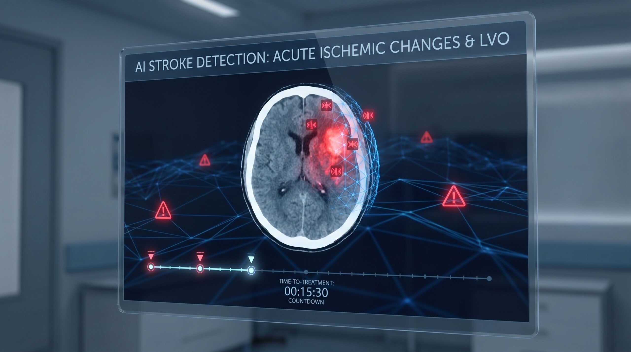
Modern AI stroke detection platforms operate as software-as-a-medical-device (SaMD) solutions that sit atop existing imaging infrastructure. When a patient with suspected stroke undergoes non-contrast CT (NCCT) and CT angiography (CTA), the images are automatically routed to the AI algorithm, which processes them in parallel with standard radiology review. The typical pipeline involves three key steps: automated detection of abnormal findings (LVO, hemorrhage, or perfusion deficit), quantification of the extent and location of pathology, and instant notification of the stroke team via mobile alert with annotated images.
CT Angiography–Based LVO Detection
The majority of FDA-cleared stroke AI platforms focus on automated detection of anterior circulation large vessel occlusions on CTA. Deep learning algorithms — typically convolutional neural networks trained on thousands of annotated CTA datasets — identify occlusions in the internal carotid artery (ICA) and M1 segment of the middle cerebral artery (MCA). Reported sensitivity ranges from 82% to 97% across platforms, with specificity generally between 72% and 98%. For example, the Viz-AI Algorithm v4.1.2 achieved 82% sensitivity and 94% specificity for proximal LVO detection, while RAPID-LVO (RapidAI) reported 96% sensitivity and 98% specificity in validation studies.
However, detection of more distal occlusions — M2 segment MCA and posterior circulation vessels — remains a significant challenge. Published sensitivities for M2 occlusion detection range from only 49% to 72% across current commercial platforms. A 2025 head-to-head study of 1,122 code stroke patients presented at the European Stroke Organisation Conference (ESOC) found that RapidAI’s CT perfusion software detected 93% of medium vessel occlusions (MeVOs) compared to 70% for Viz.ai’s platform, illustrating meaningful performance differences between systems. This is a critical frontier, as emerging evidence supports expanded indications for thrombectomy in selected MeVO patients.
CT Perfusion Automated Analysis
Alongside CTA-based detection, automated CT perfusion (CTP) analysis has become integral to stroke triage — particularly for determining treatment eligibility in the extended time window (6–24 hours from symptom onset). RapidAI’s RAPID CTP module, used in the pivotal DAWN and DEFUSE 3 trials, automatically generates ischemic core and penumbra maps, enabling rapid identification of patients with salvageable brain tissue. These automated perfusion maps have been incorporated into both AHA/ASA and ESO guidelines as decision-support tools for late-presenting stroke patients.
The integration of CTP with LVO detection creates a comprehensive triage package: the AI simultaneously identifies the occlusion site, quantifies the volume of irreversibly injured tissue, and estimates the amount of brain at risk — providing the stroke team with actionable information within minutes of scan completion. This multimodal approach is increasingly regarded as the standard for AI-enhanced stroke imaging, as reflected in foundation model architectures that aim to unify multiple imaging inputs into a single diagnostic framework.
Clinical Evidence: Workflow Gains and the Outcomes Gap
Treatment Time Reductions
The strongest evidence for AI stroke platforms centers on workflow acceleration. A 2025 systematic review and meta-analysis published in Translational Stroke Research pooled data from 12 studies encompassing 15,595 patients to evaluate the impact of Viz.ai implementation on stroke workflow metrics. The analysis demonstrated significant reductions in CT-to-EVT time (standardized mean difference −0.71, p < 0.001), door-to-groin puncture time (SMD −0.50, p < 0.001), and CT-to-recanalization time (SMD −0.55, p < 0.001). Door-in door-out times at spoke hospitals were also significantly shorter in the post-AI cohort (SMD −0.49, p < 0.001).
Separately, a multicenter retrospective analysis of 474 patients (215 post-Viz implementation) presented at the International Stroke Conference (ISC) 2025 reported that Viz LVO implementation reduced treatment time by an average of 31 minutes. The investigators noted that the platform’s impact extends beyond algorithmic detection alone — the integrated care coordination features, including secure mobile image sharing, real-time dashboards, and automated team activation, contribute substantially to the observed workflow improvements.
The Clinical Outcomes Question
Despite consistent and clinically meaningful reductions in treatment times, the translation of these gains into improved patient outcomes has proven elusive. The same 2025 meta-analysis that documented significant workflow improvements found no statistically significant differences in clinical outcomes between pre- and post-AI implementation groups — including 90-day modified Rankin Scale (mRS) scores ≤ 2, symptomatic intracranial hemorrhage, mortality, and length of hospital stay (all p > 0.05).
This paradox — faster treatment without detectable outcome improvement — likely reflects several confounding factors. Most studies are pre-post observational designs with significant intervals between cohorts, during which multiple other stroke workflow modifications may have occurred. Furthermore, many variables beyond the AI platform’s control influence patient outcomes, including presentation severity, comorbidities, procedural technique, and post-procedural care. The absence of randomized controlled trial data specifically evaluating AI stroke platforms remains the most significant evidence gap in this field.
| Platform | LVO Sensitivity | LVO Specificity | Key Capabilities | Hospital Deployment |
|---|---|---|---|---|
| Viz.ai (Viz LVO) | 82–90% | 82–94% | LVO detection, mobile alerts, care coordination, 50+ FDA-cleared algorithms | 1,700+ hospitals (US + Europe) |
| RapidAI (RAPID LVO/CTP) | 93–96% | 90–98% | LVO/MeVO detection, CTP perfusion maps, ASPECTS scoring, 5 new 2025 FDA clearances | 2,300+ hospitals globally |
| Brainomix (e-Stroke) | 84% | 96% | LVO detection, e-CTA, e-ASPECTS, NCCT hemorrhage, ESO-endorsed | 400+ hospitals (Europe-focused) |
| Aidoc | ~90% | ~91% | ICH and LVO triage, CARE1 foundation model (FDA cleared Feb 2025) | 1,000+ hospitals |
| Methinks AI (NCCT Stroke) | Pending publication | Pending publication | LVO + hemorrhage detection on non-contrast CT only; CE-marked + FDA cleared 2025 | Early deployment |
AI scan-to-notification time vs. 15–60 min traditional workflow
Average treatment time reduction with Viz.ai (ISC 2025, n=474)
Sensitivity range for LVO detection across FDA-cleared platforms
US stroke hospitals using AI as of 2025; only 12% in rural settings
Beyond LVO: Hemorrhage Detection and Emerging Applications
While LVO detection dominates the AI stroke landscape, several platforms have expanded their capabilities to include automated detection of intracerebral hemorrhage (ICH) on non-contrast CT. AI-based ICH detection generally achieves higher accuracy than LVO identification, as acute hemorrhage produces stark contrast on NCCT that is well-suited to pattern recognition. A multicenter study of Aidoc’s hemorrhage detection system across 12 hospitals demonstrated strong sensitivity, and the platform’s triage function has been shown to significantly reduce time-to-radiologist-notification for positive cases.
Methinks AI achieved a notable milestone in 2025 by becoming the first company to receive both CE marking and FDA 510(k) clearance for AI-based LVO detection on non-contrast CT alone — eliminating the requirement for contrast-enhanced CTA. This development has particular implications for community hospitals and resource-limited settings where CTA may not be immediately available, potentially democratizing access to AI-assisted stroke triage. Additionally, emerging platforms are exploring AI-powered detection of posterior circulation strokes, automated ASPECTS scoring for infarct extent quantification, and prediction of hemorrhagic transformation risk following thrombolysis — areas where current tools have notable limitations.
Implementation Challenges: From Algorithm to Impact
Hub-and-Spoke Network Integration
The real-world impact of AI stroke detection extends far beyond algorithm accuracy. In hub-and-spoke stroke networks — where primary stroke centers (PSCs) transfer eligible patients to comprehensive stroke centers (CSCs) for thrombectomy — AI platforms serve as critical coordination tools. By enabling real-time image sharing between referring and receiving hospitals, these systems allow neurointerventionalists to review imaging and make transfer decisions before the patient arrives, facilitating parallel rather than sequential workflows. A 2025 economic analysis presented at ISC found that AI-assisted coordination reduced futile transfers and produced significant financial benefits for PSC centers by ensuring patients receive care at the appropriate level.
However, the platforms function as comprehensive workflow tools rather than standalone diagnostic algorithms. Viz.ai’s impact on stroke metrics, for example, is likely multifactorial: the deep learning LVO detection is one component, but the automated alerts, secure mobile communication, real-time dashboards, and streamlined team activation contribute substantially to the observed improvements. This distinction is important when evaluating AI stroke platforms — workflow integration may matter as much as, or more than, raw diagnostic accuracy.
Adoption Barriers and Health Equity
Despite the compelling evidence for workflow improvement, AI adoption in stroke care remains limited. As of 2025, only an estimated 20–30% of US hospitals with stroke programs have implemented AI detection systems, with deployment concentrated in larger urban centers. Rural hospitals — which arguably stand to benefit most from AI-assisted triage given their limited access to subspecialty neuroradiology — have the lowest adoption rates, estimated at approximately 12%. Cost is a primary barrier, with annual licensing fees for AI stroke platforms ranging from $50,000 to $250,000, compounded by IT infrastructure requirements and the need for staff training.
This adoption pattern raises significant health equity concerns. Black and Hispanic populations in the United States experience higher stroke incidence, greater severity, and worse outcomes compared to White patients — disparities driven by socioeconomic factors, comorbidity burden, and differential access to specialized stroke care. If AI tools remain concentrated in well-resourced centers, they risk widening rather than narrowing existing disparities. Addressing this challenge will require innovative pricing models, cloud-based deployment options that reduce infrastructure requirements, and policy interventions to incentivize AI adoption in underserved communities. The evolving role of radiologists in the AI era includes advocacy for equitable technology deployment.
NCCT + CTA ± CTP acquired at presenting hospital. Images auto-routed to AI platform in parallel with PACS.
Algorithm detects LVO/ICH, generates perfusion maps, calculates ASPECTS. Confidence score and annotated images produced.
Mobile alert to stroke neurologist + neurointerventionalist with images. Parallel activation of IR suite, anesthesia, and transfer team.
Physician reviews AI output + clinical context. EVT/thrombolysis decision made. If transfer needed, receiving center already has images.
Future Directions
Several developments are poised to reshape AI-assisted stroke care over the next two to three years. First, the expansion of AI detection capabilities to medium vessel occlusions and posterior circulation strokes will address the most significant diagnostic blind spots of current systems. Emerging algorithms trained specifically on M2/M3 occlusions and basilar artery thrombosis show promising early results, though clinical validation is ongoing.
Second, the emergence of foundation models in medical imaging — large, pre-trained models capable of analyzing multiple imaging modalities and clinical data simultaneously — offers the potential for truly multimodal stroke triage. Rather than separate algorithms for LVO detection, perfusion analysis, and hemorrhage screening, a unified foundation model could integrate NCCT, CTA, and CTP data with clinical variables (age, NIHSS score, onset time) to provide a comprehensive treatment recommendation. Aidoc’s CARE1 foundation model, which received FDA clearance in February 2025, represents the first step in this direction.
Third, advances in portable and point-of-care imaging — exemplified by Hyperfine’s AI-powered portable MRI system, which received FDA clearance for enhanced stroke DWI in December 2025 — may enable AI-assisted stroke detection in prehospital settings, emergency departments without CT capability, and low-resource environments. The combination of portable imaging hardware with cloud-based AI analysis could fundamentally alter the geography of stroke care.
Finally, the generation of prospective, randomized evidence evaluating the impact of AI on patient outcomes — not just workflow metrics — remains the field’s most pressing priority. Without such data, the widespread adoption and reimbursement of AI stroke tools will continue to face barriers. The development of a dedicated Medicare reimbursement pathway for AI diagnostic devices, currently under congressional consideration, may help align incentives with evidence generation.
Clinical Implications
The evidence supporting AI-assisted stroke detection and triage is substantial for workflow optimization and increasingly mature for diagnostic accuracy, but it remains incomplete for the outcome metric that matters most: whether AI improves how patients actually do after stroke. The consistent finding of 30–52% reductions in treatment delays is clinically meaningful in a disease where “time is brain,” yet the failure to demonstrate statistically significant outcome improvement in pooled analyses should temper uncritical enthusiasm.
For stroke programs considering AI implementation, the current evidence supports adoption as a workflow enhancement tool — particularly for hub-and-spoke networks, hospitals with limited neuroradiology coverage, and centers seeking to reduce treatment variability. However, the choice of platform matters: head-to-head comparisons reveal meaningful differences in detection sensitivity, particularly for MeVOs and posterior circulation strokes. Clinicians should evaluate platforms not only on LVO detection accuracy but on the comprehensiveness of the care coordination ecosystem, integration with existing IT infrastructure, and the strength of real-world validation data. In all cases, AI serves as a triage and notification tool — the final diagnostic and treatment decision remains with the physician, as mandated by current FDA classification of these systems as Class II devices requiring human oversight.
References
- Sarhan K, Azzam AY, Moawad MHE, et al. Automated emergent large vessel occlusion detection using Viz.ai software and its impact on stroke workflow metrics and patient outcomes in stroke centers: a systematic review and meta-analysis. Transl Stroke Res. 2025;16:2258-2271. doi:10.1007/s12975-025-01354-0
- Siegler JE, et al. Treatment time metrics following implementation of the Viz.ai artificial intelligence intracranial occlusion-detection and communication platform: a multicenter retrospective analysis. Presented at: ISC 2025; February 5-7, 2025; Los Angeles, CA.
- Sachdev H, Hudson A, Ong K, et al. AI detection of medium vessel occlusions: evaluating performance of RapidAI vs. Viz.ai CT perfusion in 1,591 consecutive code strokes. Presented at: ESOC 2025; May 21-23, 2025; Helsinki, Finland.
- Goyal M, Menon BK, van Zwam WH, et al. Endovascular thrombectomy after large-vessel ischaemic stroke: a meta-analysis of individual patient data from five randomised trials. Lancet. 2016;387(10029):1723-1731.
- Nogueira RG, Jadhav AP, Haussen DC, et al. Thrombectomy 6 to 24 hours after stroke with a mismatch between deficit and infarct (DAWN). N Engl J Med. 2018;378(1):11-21.
- Albers GW, Marks MP, Kemp S, et al. Thrombectomy for stroke at 6 to 16 hours with selection by perfusion imaging (DEFUSE 3). N Engl J Med. 2018;378(8):708-718.
- Saver JL, Goyal M, van der Lugt A, et al. Time to treatment with endovascular thrombectomy and outcomes from ischemic stroke: a meta-analysis. JAMA. 2016;316(12):1279-1288.
- Dantas J, Barros G, Mutarelli A, et al. Detection of large vessel occlusion using artificial intelligence tools: a systematic review and meta-analysis. Neuroradiology. 2025;67:2399-2407. doi:10.1007/s00234-025-03716-9
- Nagaratnam K, Mathieson P, Podlasek A, et al. Artificial intelligence assisted detection of large vessel occlusion on CT angiography in acute stroke patients: a multi-reader multi-case study. BJR Artif Intell. 2025;2(1):ubaf001.
- Slater LA, et al. RapidAI compared with human readers of acute stroke imaging for detection of intracranial vessel occlusion. Stroke Vasc Interv Neurol. 2024;4(3):e001145.
- Viz.ai. Two new studies demonstrate proven impact of Viz.ai’s stroke solution on patient outcomes and hospital economics. Press release. February 13, 2025.
- Mortin M, Shifflett B, Meyer DM, et al. CONNECT: Coordinating options for neurovascular patients needing electrophysiology consults and treatments. Neurohospitalist. 2025. Published online ahead of print.
- RapidAI. FDA clearance of five new imaging modules: Rapid DeltaFuse, Rapid LMVO, Rapid MLS, Rapid OH, Rapid Aortic. Press release. November 25, 2025.
- US Food and Drug Administration. Artificial intelligence and machine learning (AI/ML)-enabled medical devices. FDA Device Tracker. Updated January 2026.
- Powers WJ, Rabinstein AA, Ackerson T, et al. Guidelines for the early management of patients with acute ischemic stroke: 2019 update. Stroke. 2019;50(12):e344-e418.
- Turc G, Bhogal P, Fischer U, et al. European Stroke Organisation (ESO) – European Society for Minimally Invasive Neurological Therapy (ESMINT) guidelines on mechanical thrombectomy in acute ischaemic stroke. Eur Stroke J. 2023;8(4):802-832.
- Hyperfine Inc. FDA clearance of Optive AI software update with advanced diffusion imaging capability for stroke detection with Swoop system. Press release. December 15, 2025.
- Elhawary T, Mansour Y, Abdelrazek O, et al. Current stroke solutions using artificial intelligence: a review of the literature. J Clin Med. 2024;13(24):7797. doi:10.3390/jcm13247797
Disclaimer: This article is intended for educational purposes and does not constitute medical advice. Clinical decisions should be based on individual patient assessment and current institutional protocols. MedTrainHub has no financial relationship with any AI platform manufacturer mentioned in this article.
Conflicts of Interest: None declared.
Suggested Citation: MedTrainHub Editorial Team. AI-Assisted Stroke Detection and Triage: From Scan to Treatment in Minutes. MedTrainHub.com. April 2026.