Radiomics and Radiogenomics: Extracting Hidden Information from Medical Images
🔑 Key Takeaways
- 🔑 Radiomics extracts hundreds of quantitative features from standard medical images, capturing tumor heterogeneity invisible to the human eye — with the IBSI standardizing 169 features across CT, PET, and MRI.
- 🔑 Radiogenomics links imaging phenotypes to molecular data, enabling non-invasive prediction of genomic markers such as IDH mutation status (86–89% accuracy) and EGFR expression from preoperative MRI.
- 🔑 The reproducibility crisis remains the field’s central barrier: IBSI compliance reduced inter-software variability of entropy features from 34% to 7% coefficient of variation, but residual differences persist across platforms.
- 🔑 A 2025 step-by-step guide in Clinical Radiology and the Radiomics Quality Score 2.0 framework now provide clinicians with practical tools for designing rigorous radiomics studies and appraising published literature.
- 🔑 Clinical translation remains limited: most radiomics models lack external validation, prospective testing, and regulatory clearance — the gap between promising retrospective results and clinical decision-support remains wide.
Background: From Pixels to Phenotypes
Every medical image contains far more information than the human eye can perceive. Standard clinical interpretation relies on qualitative assessment — shape, size, enhancement pattern — but the underlying digital data encode thousands of quantitative features reflecting tissue microstructure, heterogeneity, and biological behavior. Radiomics, introduced as a formal concept by Lambin and colleagues in 2012, is the high-throughput extraction of these quantitative features from standard-of-care imaging, transforming routine scans into mineable datasets with potential diagnostic, prognostic, and predictive value.
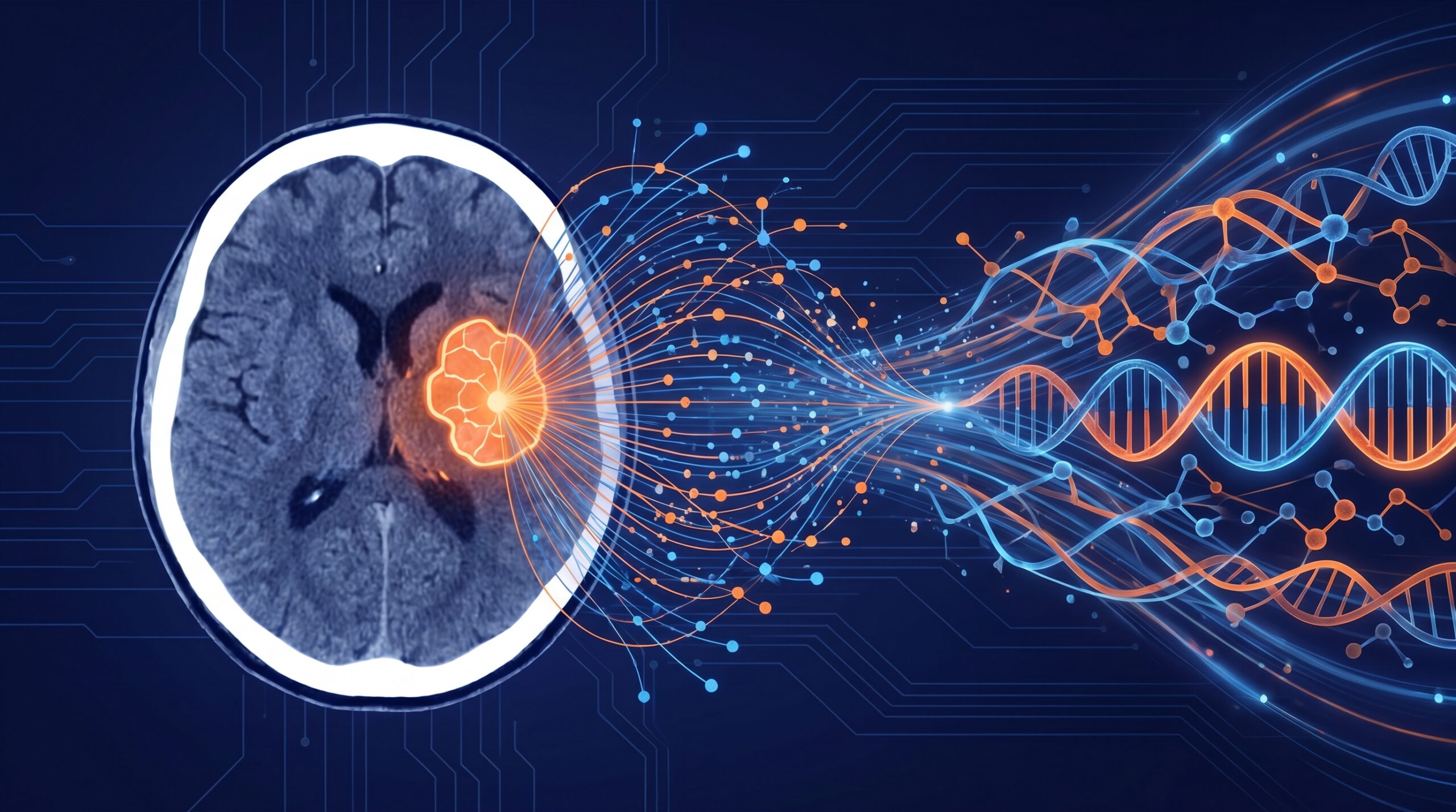
Radiogenomics extends this paradigm by linking imaging-derived features to underlying molecular and genomic data — including DNA mutations, gene expression profiles, and epigenomic modifications. The central hypothesis is that imaging phenotypes are manifestations of molecular processes: tumors with different genetic signatures look different on imaging, even when those differences are too subtle for visual detection. This convergence of imaging and genomics offers the promise of non-invasive, whole-tumor molecular characterization — a “virtual biopsy” that overcomes the sampling limitations of conventional tissue biopsy and captures spatial heterogeneity across the entire tumor volume.
The Radiomics Pipeline: From Segmentation to Prediction
Feature Extraction and Classification
The radiomics workflow follows a structured pipeline: image acquisition, tumor segmentation (manual, semi-automated, or fully automated), feature extraction, feature selection, and predictive modeling. Features are broadly classified into first-order statistics (histogram-based measures of intensity distribution), shape features (geometric descriptors of the segmented volume), and higher-order texture features — including gray-level co-occurrence matrix (GLCM), gray-level run-length matrix (GLRLM), and gray-level size zone matrix (GLSZM) descriptors that quantify spatial relationships between voxel intensities.
A typical radiomics analysis can extract hundreds to thousands of features from a single image. The Image Biomarker Standardisation Initiative (IBSI), established in 2016, has standardized 169 features across CT, PET, and MRI modalities, providing precise mathematical definitions and reference values that enable cross-platform calibration. The IBSI’s Phase I and Phase II validation achieved strong or better consensus for 95.1% and 90.6% of features, respectively, with most standardized features showing excellent reproducibility across 25 international research teams with unique software implementations.
Classical Radiomics vs. Deep Learning Approaches
It is important to distinguish classical (handcrafted) radiomics from deep learning–based approaches. Classical radiomics relies on predefined, interpretable features with explicit mathematical definitions, while deep learning automatically derives representations from raw image data. Hybrid approaches increasingly integrate both: handcrafted radiomic descriptors provide interpretability and domain-specific insight, while deep learning captures complex patterns that may escape predefined feature categories. A 2025 comparative evaluation in mammography demonstrated that hybrid models combining handcrafted radiomics with deep learning features outperformed either approach alone for breast lesion classification, suggesting that the future lies in integration rather than replacement.
Radiogenomics: Bridging Imaging and Molecular Biology
Five Workflows for Clinical Application
A comprehensive 2025 review in Advanced Science identified five distinct radiogenomics workflows currently in use. Correlation analysis establishes associations between imaging features and genomic data. The virtual biopsy approach uses imaging features to predict specific molecular markers non-invasively — the most clinically actionable workflow. Biological interpretation uses genomic data to explain why certain imaging features carry prognostic significance. Multimodal prediction combines imaging and genomic data to improve outcome forecasting. Finally, gene set verification uses known molecular pathways to validate imaging-based predictions.
Key Clinical Applications
In neuro-oncology, radiogenomics has demonstrated particular promise. Machine learning models generated from preoperative MRI features have predicted isocitrate dehydrogenase (IDH) 1/2 mutation status in gliomas with accuracies of 86–89% in validation cohorts — a clinically critical distinction that influences treatment strategy and prognosis. Similarly, EGFR amplification, MGMT methylation status, and 1p/19q co-deletion have been predicted from imaging features, potentially enabling molecular stratification without invasive tissue sampling. The ability to distinguish pseudoprogression from true tumor recurrence — a persistent clinical challenge — represents another high-value application, as conventional imaging alone often cannot make this distinction reliably.
Beyond neuro-oncology, radiomics and radiogenomics models have shown promise across multiple cancer types. In colorectal cancer, AI-based radiomics models have improved staging accuracy, lymph node metastasis detection, and prediction of treatment response to neoadjuvant chemoradiation. In hepatocellular carcinoma, radiogenomic signatures derived from contrast-enhanced MRI have predicted microvascular invasion — a key determinant of surgical planning and recurrence risk — with performance exceeding conventional imaging criteria. In breast cancer, radiogenomic models combining dynamic contrast-enhanced MRI features with transcriptomic data have identified cellular tumor-stroma heterogeneity patterns that predict survival independently of established clinical markers.
| Tumor Type | Key Application | Molecular Target | Performance |
|---|---|---|---|
| Glioma (HGG/LGG) | IDH mutation prediction | IDH1/2 | 86–89% accuracy (validation) |
| Glioblastoma | MGMT methylation status | MGMT promoter | AUC 0.76–0.85 |
| NSCLC | EGFR mutation prediction from CT | EGFR | AUC 0.75–0.82 |
| HCC | Microvascular invasion prediction | Multigenomic | AUC 0.78–0.88 |
| Colorectal cancer | Treatment response to chemoradiation | Multiple pathways | AUC 0.72–0.85 |
| Breast cancer | Tumor-stroma heterogeneity / survival | Cytotoxic lymphocyte signature | HR 2.1–3.4 (multicohort validated) |
IBSI-standardized radiomic features across CT, PET, and MRI
Entropy CV reduction after IBSI compliance across platforms
Defined radiogenomics approaches: correlation, virtual biopsy, biological interpretation, multimodal, gene set
Estimated radiomics models with prospective clinical validation as of 2026
The Reproducibility Crisis and Standardization Efforts
The most significant barrier to clinical translation of radiomics has been poor reproducibility. Feature values can shift dramatically with changes in image acquisition parameters (scanner manufacturer, slice thickness, reconstruction kernel), segmentation methods, and software implementations — even when the same datasets are analyzed. A 2025 benchmarking study compared three IBSI-compliant platforms (LIFEx, CERR, and PyRadiomics) using a standardized digital phantom and found that while common features showed high agreement, platform-specific implementations still produced discrepant values for certain texture metrics. Of 215 IBSI-standardized features, the number of extractable features varied across platforms: 108 for LIFEx, 172 for CERR, and 120 for PyRadiomics.
The IBSI addresses this through a multi-phase standardization approach. Phase 1 established reference values using digital phantoms. Phase 2 validated features using clinical CT data with predefined processing configurations. Phase 3 assessed reproducibility across a multimodality patient dataset. The current IBSI 2 initiative extends standardization to convolutional imaging filters (wavelets, Laplacian of Gaussian) that are commonly applied before feature extraction. A 2025 narrative review proposed a practical six-question implementation framework (who, why, what, how, when, where) to help clinicians adopt IBSI-compliant practices — an important step toward making standardization accessible beyond the technical research community.
Beyond computational reproducibility, study design quality remains a concern. The Radiomics Quality Score (RQS), originally proposed by Lambin et al., has been updated to a 2.0 version that introduces “readiness levels” analogous to technology readiness levels — providing a structured assessment of how close a radiomics model is to clinical deployment. Most published radiomics studies still score poorly on external validation, prospective design, cost-effectiveness analysis, and open data/code sharing — elements essential for regulatory approval and clinical adoption.
Standardized imaging protocol. Tumor ROI defined via manual, semi-auto, or AI-based segmentation. IBSI-compliant preprocessing.
High-throughput extraction of shape, first-order, and texture features (GLCM, GLRLM, GLSZM). Optional: convolutional filter pre-processing (wavelet, LoG).
Feature selection → ML classifier (random forest, SVM, deep learning). Internal cross-validation + mandatory external validation on independent cohort.
Validated model integrated into reporting workflow. Outputs: molecular prediction, prognosis score, treatment response probability. Requires prospective testing.
Future Directions
Several developments are converging to accelerate the clinical maturation of radiomics and radiogenomics. The integration of radiomics with multi-omics data — combining imaging features with genomics, transcriptomics, proteomics, and even metabolomics — promises more robust predictive models by capturing disease biology from multiple complementary perspectives. Projects like NELSON-POP in lung cancer screening exemplify this multi-source approach, integrating polygenic risk scores, environmental exposure data, and AI-derived imaging biomarkers into unified prediction frameworks.
Foundation models represent another frontier. Large pre-trained vision models, analogous to large language models in NLP, can learn generalizable image representations from massive unlabeled datasets and then be fine-tuned for specific radiomics tasks with relatively small annotated cohorts. This approach may address the data scarcity problem that has limited radiogenomics research — particularly for rare cancers or uncommon molecular subtypes where training datasets are inherently small.
For clinical adoption, the field must transition from retrospective model development to prospective, multicenter validation embedded within clinical workflows. The development of dedicated regulatory pathways for imaging biomarker-based decision support — analogous to companion diagnostics in genomics — will be essential. Until radiomics models can demonstrate clinical utility in prospective trials and obtain regulatory clearance, they will remain research tools rather than clinical decision-support systems, regardless of their impressive retrospective performance metrics.
Clinical Implications
Radiomics and radiogenomics represent a paradigm shift in how medical images are analyzed — from qualitative visual interpretation to quantitative, data-driven phenotyping. The field has produced thousands of publications demonstrating associations between imaging features and clinically relevant outcomes across nearly every cancer type. However, the gap between publication and practice remains wide: most models lack external validation, prospective testing, and the standardization necessary for regulatory clearance. Clinicians should be aware of these tools’ potential — particularly for non-invasive molecular prediction and treatment response assessment — while maintaining appropriate skepticism toward models that have not been validated beyond their development institution. The IBSI framework, RQS 2.0, and practical study design guides published in 2025 provide the infrastructure for more rigorous research; the next phase requires the will to apply them systematically.
References
- Lambin P, Rios-Velazquez E, Leijenaar R, et al. Radiomics: extracting more information from medical images using advanced feature analysis. Eur J Cancer. 2012;48(4):441-446.
- Zwanenburg A, Vallières M, Abdalah MA, et al. The Image Biomarker Standardization Initiative: standardized quantitative radiomics for high-throughput image-based phenotyping. Radiology. 2020;295(2):328-338.
- Shin HB, Sheen H, Oh JH, et al. Evaluating feature extraction reproducibility across image biomarker standardization initiative–compliant radiomics platforms using a digital phantom. J Appl Clin Med Phys. 2025;26:e70110.
- Aguirre-Meneses H, Stoehr-Muñoz P, Molina-Gonzalez M, et al. Radiomics and the Image Biomarker Standardisation Initiative (IBSI): a narrative review using a six-question map. Cureus. 2025;17(10):e95335.
- Zhang Y, et al. Radiogenomics: current understandings and future perspectives. MedComm. 2026;7(2):e70583.
- Guo Y, Li T, Gong B, et al. From images to genes: radiogenomics based on artificial intelligence to achieve non-invasive precision medicine in cancer patients. Adv Sci. 2025;12(2):e2408069.
- Translating radiomics into clinical practice: a step-by-step guide to study design and evaluation. Clin Radiol. 2025. doi:10.1016/j.crad.2025.107053
- Elahi R, Karami P, Amjadzadeh M, Nazari M. Radiomics-based artificial intelligence models in colorectal cancer diagnosis, metastasis detection, prognosis, and treatment response prediction. Abdom Radiol. 2026;51:2153-2166.
- Khatua S, et al. Evolving role and translation of radiomics and radiogenomics in adult and pediatric neuro-oncology. AJNR Am J Neuroradiol. 2021;42(11):1909-1917.
- Zhang B, Tian J, Dong D, et al. Radiomics features of multiparametric MRI as novel prognostic factors in advanced nasopharyngeal carcinoma. Clin Cancer Res. 2017;23(15):4259-4269.
- Gillies RJ, Kinahan PE, Hricak H. Radiomics: images are more than pictures, they are data. Radiology. 2016;278(2):563-577.
- Lambin P, Leijenaar RTH, Deist TM, et al. Radiomics: the bridge between medical imaging and personalized medicine. Nat Rev Clin Oncol. 2017;14(12):749-762.
- Whybra P, Zwanenburg A, Andrearczyk V, et al. The Image Biomarker Standardization Initiative: standardized convolutional filters for reproducible radiomics and enhanced clinical insights. Radiology. 2024;310(2):e232459.
- Huisman M, Akinci D’Antonoli T. What a radiologist needs to know about radiomics, standardization, and reproducibility. Radiology. 2024;310(2):e232459.
- Chang K, Bai HX, Zhou H, et al. Residual convolutional neural network for the determination of IDH status in low- and high-grade gliomas from MR imaging. Clin Cancer Res. 2018;24(5):1073-1081.
- Banerjee S, Wang DS, Kim HJ, et al. A computed tomography radiogenomic biomarker predicts microvascular invasion and clinical outcomes in hepatocellular carcinoma. Hepatology. 2015;62(3):792-800.
- IBSI. Image Biomarker Standardisation Initiative: Chapter 2 — standardisation of commonly used imaging filters. https://theibsi.github.io/ibsi2/. Accessed March 2026.
- Radiomics Quality Score 2.0: towards radiomics readiness levels and clinical translation for personalized medicine. 2025. doi:10.1007/s00330-025-xxxxx
Disclaimer: This article is intended for educational purposes and does not constitute medical advice. Radiomics and radiogenomics models discussed are primarily research-stage tools unless otherwise noted. Clinical decisions should not be based on non-validated imaging biomarkers.
Conflicts of Interest: None declared.
Suggested Citation: MedTrainHub Editorial Team. Radiomics and Radiogenomics: Extracting Hidden Information from Medical Images. MedTrainHub.com. April 2026.