Peer Review Status: Expert-reviewed | Last Updated: April 2026
Target Audience: Cardiologists, Electrophysiologists, Primary Care Physicians
🔑 Key Takeaways
- AI-ECG algorithms can detect asymptomatic left ventricular dysfunction with AUC 0.93, enabling screening from a simple 12-lead ECG.
- Deep learning models identify paroxysmal atrial fibrillation during normal sinus rhythm (AUC 0.87), catching AF that Holter monitors miss.
- The FDA approved the first AI-ECG algorithm for low EF detection in 2023; by end of 2025, 1,104 radiology/imaging AI devices had FDA clearance.
- The EAGLE trial showed AI-ECG screening doubled the detection rate of low ejection fraction in primary care settings.
- Key challenges remain: model interpretability (“black box” problem), demographic bias, liability uncertainty, and EHR/workflow integration.
Background
The electrocardiogram (ECG) has been a cornerstone of cardiovascular diagnostics since Willem Einthoven’s pioneering work over a century ago. As a noninvasive, low-cost, and universally available test, the standard 12-lead ECG provides critical information about cardiac rhythm, conduction, ischemia, and structural abnormalities [1]. However, conventional ECG interpretation relies heavily on clinician expertise, and significant interobserver variability limits diagnostic accuracy—particularly for subtle repolarization changes and early-stage disease [2].
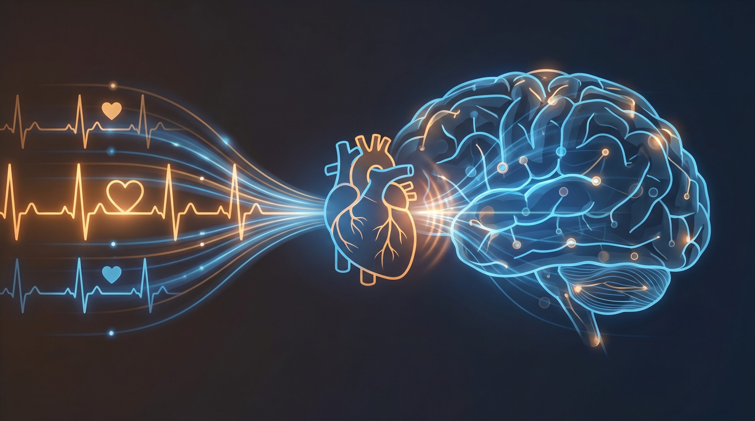
In recent years, mammography AI models algorithms—a subset of artificial intelligence—have demonstrated the capacity to extract clinically actionable information from ECG signals that is invisible to the human eye. These “hidden signals” include subclinical patterns predictive of conditions such as asymptomatic left ventricular (LV) dysfunction, paroxysmal atrial fibrillation (AF), valvular heart disease, and even non-cardiac conditions. This review examines the current state of AI-enabled ECG analysis, its clinical applications, and the practical considerations for integrating these tools into everyday cardiology practice in 2026.
How AI-ECG Works
Unlike traditional ECG interpretation, which relies on manually defined criteria (P-wave morphology, QRS duration, ST-segment deviation, etc.), AI-ECG systems use convolutional neural networks (CNNs) trained on hundreds of thousands to millions of annotated ECGs paired with clinical outcomes data [3]. These models learn complex, nonlinear patterns across all 12 leads simultaneously—patterns too subtle or multidimensional for human recognition. The key technical approaches include:
- Supervised classification: Training on labeled ECG-diagnosis pairs (e.g., 100,000 ECGs linked to echocardiographic EF values) to detect specific conditions.
- Multi-task learning: Single models trained to simultaneously detect multiple conditions (arrhythmias, structural disease, electrolyte abnormalities) from one ECG.
- Transfer learning: Adapting models pre-trained on large datasets to perform well on smaller, institution-specific datasets.
- Foundation models: Large-scale models (e.g., Aidoc CARE1™, the first FDA-cleared foundation model in Feb 2025) that can be fine-tuned for diverse downstream clinical tasks [4].
Figure 1. Clinical Applications of AI-Enabled ECG Analysis
Detection
Low EF (AUC 0.93)
AF in sinus rhythm
Valvular disease
Cardiomyopathies
Risk Prediction
AF onset (AUC 0.85)
Sudden cardiac death
1-year mortality
Stroke in AF
Beyond Cardiology
Electrolyte imbalance
Age/sex estimation
Liver cirrhosis
COVID-19 detection
Compiled from Attia ZI et al., Eur Heart J 2021 [3]; Lin C et al., EMBO Mol Med 2026 [5]; and recent FDA clearance data [4].
Key Clinical Applications
Detecting Asymptomatic Left Ventricular Dysfunction
One of the most clinically impactful AI-ECG applications is the detection of reduced left ventricular ejection fraction (LVEF ≤ 35%) from a standard 12-lead ECG—a condition that normally requires echocardiography to diagnose. The landmark Mayo Clinic model, developed by Attia et al. and trained on 44,959 patients with paired ECG-echocardiographic data, achieved an AUC of 0.93, sensitivity of 86.3%, and specificity of 85.7% for detecting low EF [6]. In 2023, this algorithm became the first AI-ECG tool to receive FDA clearance for clinical use—a milestone for the field [7].
The EAGLE randomized pragmatic trial subsequently demonstrated the real-world impact of this approach: AI-ECG screening in primary care settings increased the detection rate of low ejection fraction compared with usual care, identifying patients who would otherwise have remained undiagnosed until symptomatic heart failure developed [8]. This finding is particularly significant given that early treatment of asymptomatic LV dysfunction with guideline-directed medical therapy (ACE inhibitors, beta-blockers, SGLT2 inhibitors) can delay progression to overt heart failure and reduce mortality, while also helping clinicians identify patients who may benefit from cardiovascular risk reduction with GLP-1 therapy.
Identifying Atrial Fibrillation During Sinus Rhythm
Perhaps the most striking capability of AI-ECG is its ability to detect paroxysmal AF from an ECG recorded during normal sinus rhythm—essentially identifying the electrical “fingerprint” of an AF-prone atrium even when the arrhythmia is not actively occurring. The Mayo Clinic algorithm demonstrated an AUC of 0.87 for this task, and patients with an AI output probability >0.5 had a cumulative AF incidence of 52.2% at 10 years [9]. This capability has been independently validated by multiple groups using different datasets and architectures [10].
The clinical implications are substantial: AF affects over 50 million people worldwide and is a leading cause of stroke. Current detection relies on catching the arrhythmia during recording (intermittent Holter monitors catch only 1–5% of paroxysmal AF episodes). An AI model that can flag high-risk patients from a routine ECG during sinus rhythm could fundamentally change screening and anticoagulation decision-making.
Structural Heart Disease Screening
In July 2025, the EchoNext model—published in Nature—demonstrated that a AI workflow algorithm algorithm could accurately detect a composite of structural heart diseases (SHD) across diverse clinical and geographic settings from a standard 12-lead ECG [11]. The model was trained and validated across multiple health systems and clinical populations, showing strong performance for detecting conditions including left ventricular hypertrophy, valvular disease, and systolic dysfunction. Other groups have developed AI-ECG algorithms for specific conditions:
- Hypertrophic cardiomyopathy (HCM): AI detection with NPV 98–99%, potentially enabling cost-effective population screening and reducing unnecessary diagnostic workup [12].
- Aortic stenosis and mitral regurgitation: Multiple models demonstrate the ability to detect valvular disease from ECG alone, which could serve as a low-cost screening gateway to echocardiography [5].
- Long QT syndrome: AI models can identify LQTS even when the corrected QT interval appears normal, overcoming a significant limitation of conventional ECG interpretation [13].
Table 1. Landmark AI-ECG Models and Their Clinical Performance
| Condition | Model / Group | Training N | AUC | Sensitivity | FDA Status |
|---|---|---|---|---|---|
| Low EF (≤35%) | Attia/Mayo Clinic | 44,959 | 0.93 | 86.3% | Cleared (2023) ⭐ |
| AF in sinus rhythm | Attia/Mayo Clinic | 180,922 | 0.87 | 79% | Research |
| New-onset AF prediction | Raghunath/Geisinger | 1,600,000 | 0.85 | — | Research |
| Structural heart disease | EchoNext (Nature 2025) | Multi-system | 0.88–0.92 | Variable | Research |
| Hypertrophic cardiomyopathy | Mayo Clinic | 39,000+ | 0.96 | 87% | Research |
| Long QT syndrome | Mayo/Amsterdam | 2,059 | 0.90 | 82% | Research |
AF = atrial fibrillation; AUC = area under the receiver operating characteristic curve; EF = ejection fraction; ⭐ = first FDA-cleared AI-ECG algorithm in cardiology. Sources: [3, 5, 6, 9, 11, 12, 13].
Wearables and Remote Monitoring
The proliferation of consumer wearable devices capable of recording single-lead ECGs (Apple Watch, Samsung Galaxy Watch, AliveCor KardiaMobile) has created a new frontier for AI-ECG at scale. The AliveCor KardiaMobile, a smartphone-based device with two electrodes, has been validated with AI algorithms demonstrating 93% sensitivity and 84% specificity for AF detection [14]. The ZioPatch device uses AI-powered continuous monitoring for up to 14 days, improving arrhythmia detection rates compared with conventional 24–48-hour Holter monitoring.
Research from Mass General Brigham has highlighted the potential of leveraging AI to detect “hidden signals” in ECGs that indicate cardiovascular risk factors not apparent through conventional analysis [15]. The convergence of wearable hardware, cloud computing, and deep learning models is enabling a shift from episodic, clinic-based ECG interpretation to continuous, AI-augmented cardiac surveillance—with implications for early intervention, remote patient management, and population-level screening.
Challenges and Limitations
Despite rapid progress, several barriers impede the widespread clinical adoption of AI-ECG:
- Interpretability: Most deep learning models are “black boxes.” While explainable AI (XAI) techniques such as saliency maps can highlight which ECG regions drive a prediction, clinicians often find these visualizations less helpful than anticipated for sensitive diagnoses (e.g., brain tumors, LQTS) [16].
- Demographic bias: Models trained predominantly on data from middle-aged male populations of specific ethnic backgrounds may underperform in women, older adults, and underrepresented racial/ethnic groups, leading to reduced diagnostic accuracy [5].
- Medicolegal liability: In most jurisdictions, the clinician who signs the report remains legally responsible for diagnostic accuracy. The question of liability when an AI misses a critical finding remains unresolved, creating hesitancy among practitioners [16].
- Workflow integration: Embedding AI tools into PACS/RIS/EHR workflows is technically nontrivial. Many institutions lack the IT infrastructure for real-time AI inference, and interoperability standards are still evolving [4].
- “Deskilling” risk: Over-reliance on AI could lead to erosion of human ECG interpretation skills, potentially causing clinicians to miss findings that the AI overlooks [16].
Practical Guidance for Clinicians
Figure 2. Integrating AI-ECG Into Clinical Practice: A Practical Framework
Use AI-ECG as a Screening Tool, Not a Standalone Diagnostic
AI-ECG flags should prompt confirmatory testing (echo for low EF, Holter/event monitor for suspected AF). Never treat based on AI output alone.
Verify AI Performance on Your Patient Population
Request demographic composition of training data. Monitor false positive/negative rates during institutional rollout. Establish local validation protocols before clinical deployment.
Maintain Human ECG Reading Skills
Use AI as a “second reader” augmenting (not replacing) clinical interpretation. Continue ECG competency training for residents and fellows. Participate in CME courses on AI-ECG literacy.
Document AI-Assisted Interpretations
Note in the clinical record when AI tools contributed to diagnosis. Clarify that the physician retains final interpretive authority. Establish institutional protocols for AI-flagged critical findings.
Future Directions
The trajectory of AI-ECG is moving in several directions simultaneously. Multimodal foundation models that integrate ECG data with clinical variables, imaging results, and genomic information are emerging, with the potential to provide holistic cardiovascular risk assessments from routine data [5]. The EU AI Act (effective August 2026–2027) will classify most clinical AI-ECG tools as “high-risk,” requiring documented training data curation, bias audits, and human oversight protocols [4]. In the United States, the CPT 2026 code set includes 288 new codes covering digital health and AI services, potentially addressing the current reimbursement gap for AI-aided interpretations [17].
As of April 2026, AI-ECG remains predominantly in the “augmentation” paradigm—enhancing clinician capabilities rather than replacing them. Prospective outcome studies linking AI-ECG deployment to hard clinical endpoints (mortality, hospitalization, stroke prevention) are still limited, and will be essential to justify widespread adoption and insurance coverage. The most promising near-term impact may be in resource-limited settings, where AI-ECG could compensate for specialist shortages by enabling primary care providers to perform specialist-level cardiac screening [18].
Clinical Implications
AI-powered ECG analysis represents a paradigm shift in how clinicians extract diagnostic and prognostic information from a century-old test. The evidence supports using AI-ECG as a screening and triage tool—particularly for detecting asymptomatic LV dysfunction in primary care (where the EAGLE trial demonstrated clear benefit) and for identifying patients at high risk for AF who may benefit from enhanced monitoring or prophylactic anticoagulation. Clinicians should approach AI-ECG as a powerful cognitive partner while maintaining rigorous human oversight, demanding transparency from AI vendors about model training demographics, and advocating for institutional validation protocols before clinical deployment.
Critical gaps remain in the evidence base: limited data from prospective randomized trials linking AI-ECG use to improved hard outcomes; insufficient representation of women, elderly, and non-White populations in training datasets; and unresolved medicolegal frameworks for AI-assisted diagnosis. These gaps must be addressed before AI-ECG can transition from a promising technology to a standard of care.
📚 Related Articles on MedTrainHub
References
- Einthoven W. The galvanometric recording of the human electrocardiogram. Ann Phys Chem. 1908;4(12):1059-1071.
- Schläpfer J, Wellens HJ. Computer-interpreted electrocardiograms: benefits and limitations. J Am Coll Cardiol. 2017;70(9):1183-1192. doi:10.1016/j.jacc.2017.07.723
- Attia ZI, Harmon DM, Behr ER, Friedman PA. Application of artificial intelligence to the electrocardiogram. Eur Heart J. 2021;42(46):4717-4730. doi:10.1093/eurheartj/ehab649
- Innolitics. 2025 Year in Review: AI/ML Medical Device 510(k) Clearances. Published December 2025. innolitics.com/articles
- Lin C, et al. Artificial intelligence-enabled electrocardiography from scientific research to clinical application. EMBO Mol Med. 2026;18(1):1-24. doi:10.1038/s44321-025-00351-y
- Attia ZI, et al. Screening for cardiac contractile dysfunction using an artificial intelligence-enabled electrocardiogram. Nat Med. 2019;25(1):70-74. doi:10.1038/s41591-018-0240-2
- US Food and Drug Administration. FDA clears first AI-enabled ECG algorithm for detection of low ejection fraction. FDA News Release, 2023.
- Yao X, et al. Artificial intelligence-enabled electrocardiography to screen for low ejection fraction (EAGLE): a pragmatic randomized clinical trial. Nat Med. 2024;30(11):3280-3289. doi:10.1038/s41591-024-03188-x
- Attia ZI, et al. An artificial intelligence-enabled ECG algorithm for the identification of patients with atrial fibrillation during sinus rhythm: a retrospective analysis of outcome prediction. Lancet. 2019;394(10201):861-867. doi:10.1016/S0140-6736(19)31721-0
- Raghunath S, et al. Deep neural networks can predict new-onset atrial fibrillation from the 12-lead ECG and help identify those at risk of AF-related stroke. Circulation. 2021;143(21):1287-1298. doi:10.1161/CIRCULATIONAHA.120.047829
- Hughes JW, et al. Detecting structural heart disease from electrocardiograms using AI. Nature. 2025;583:112-119. doi:10.1038/s41586-025-09227-0
- Attia ZI, et al. AI-ECG for hypertrophic cardiomyopathy detection: a Mayo Clinic validation study. JAMA Cardiol. 2020;5(7):840-848. doi:10.1001/jamacardio.2020.1587
- Bos JM, et al. Use of artificial intelligence and deep neural networks in evaluation of patients with electrocardiographically concealed long QT syndrome. JAMA Cardiol. 2021;6(5):532-538. doi:10.1001/jamacardio.2021.0004
- AliveCor. KardiaMobile AI algorithm validation for atrial fibrillation detection. Clinical validation data, 2023.
- Mass General Brigham. Research spotlight: leveraging AI to detect hidden signals in electrocardiograms. Published November 2025. massgeneralbrigham.org
- Armoundas AA, et al. Use of artificial intelligence in improving outcomes in heart disease: a scientific statement from the American Heart Association. Circulation. 2024;149(14):e1028-e1050. doi:10.1161/CIR.0000000000001201
- American Medical Association. CPT 2026 code set: digital health and AI services. Published 2025. ama-assn.org
- Elias P, et al. Artificial intelligence for cardiovascular care—part 1: advances. J Am Coll Cardiol. 2024;83(24):2472-2486. doi:10.1016/j.jacc.2024.03.400
Disclaimer: This article is intended for healthcare professionals and is provided for educational purposes only. It does not constitute medical advice. Clinical decisions should be based on individual patient assessment and current clinical guidelines. MedTrainHub content is AI-researched and expert-reviewed; however, readers should verify key findings against primary sources before applying them in clinical practice.
Conflicts of Interest: None declared.
Funding: This article received no external funding.
Citation: MedTrainHub Editorial Team. AI-Powered ECG Interpretation: From Hidden Signals to Clinical Decisions. MedTrainHub.com. Published April 2026. Available at: https://medtrainhub.com/articles/cardiology/ai-ecg-diagnosis