Peer Review Status: Expert-reviewed | Last Updated: April 2026
Target Audience: Radiologists, Imaging Specialists, Radiology Administrators
🔑 Key Takeaways
- The FDA has authorized 1,451 AI-enabled medical devices through end-2025; 1,104 (76%) are radiology devices.
- In 2025 alone, 295 new AI/ML devices received FDA clearance — 62% were Software as a Medical Device (SaMD).
- Aidoc’s CARE1™ became the first FDA-cleared foundation model in clinical AI (February 2025), marking a new era for adaptive radiology tools.
- Deep learning MRI reconstruction can reduce scan times by up to 50% while maintaining or improving image quality.
- The dominant paradigm is augmentation, not replacement: AI serves as a cognitive partner enhancing reproducibility, efficiency, and quantitative rigor.
Background
Radiology was among the first medical specialties to fully digitize, and it now stands at the forefront of AI integration in healthcare. The application of deep learning to foundation models in radiology AI has evolved from early proof-of-concept studies into a rapidly maturing clinical infrastructure. As of late 2025, the FDA had authorized 1,451 AI-enabled medical devices, of which 1,104 (76%) were radiology-specific—underscoring medical imaging’s dominant position in the clinical AI landscape [1, 2]. These tools now assist radiologists in image interpretation, workflow triage, quantitative analysis, and even preliminary report generation.

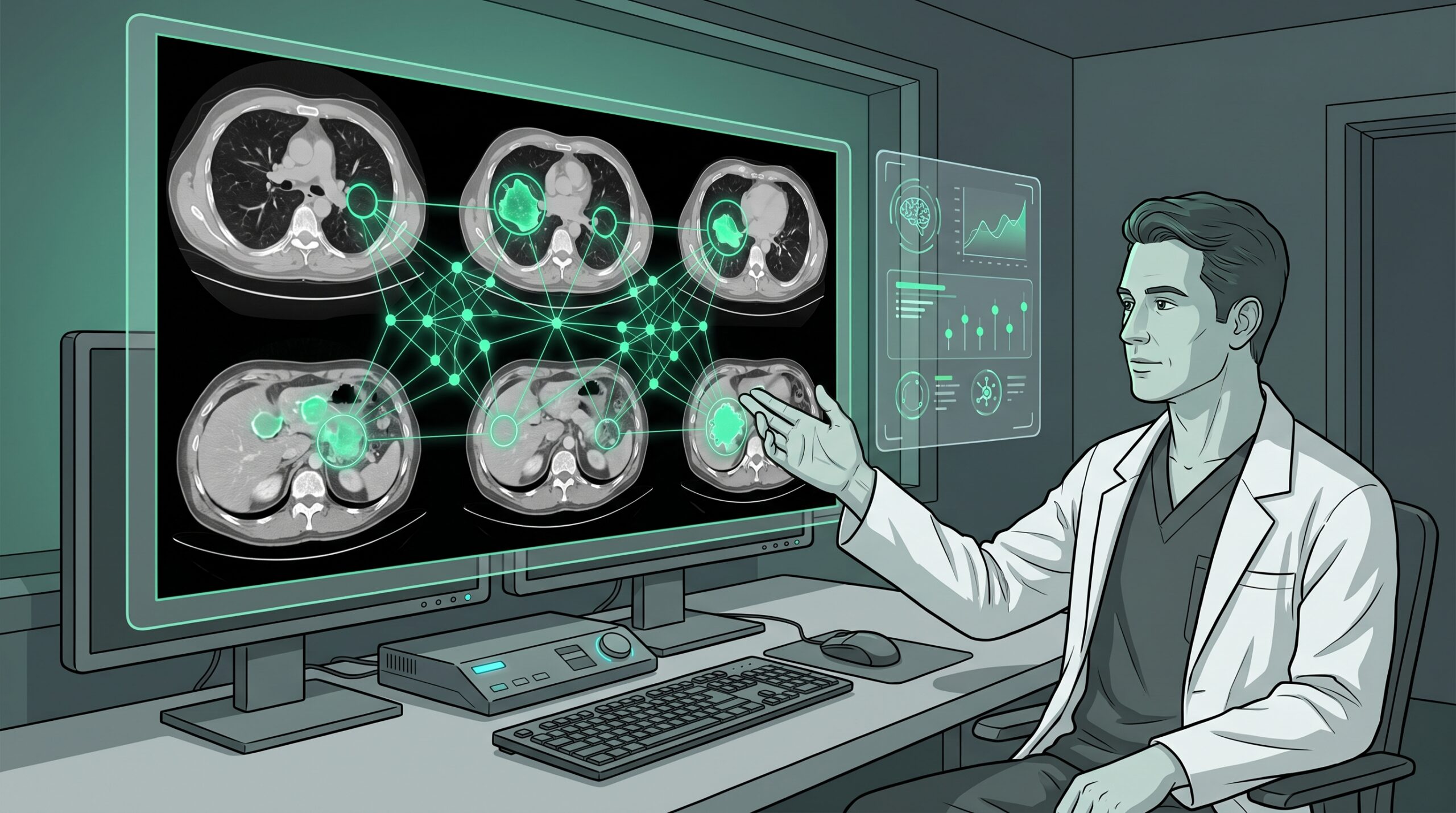
The 2025 annual meeting of the Radiological Society of North America (RSNA) and the 2026 European Congress of Radiology (ECR) both confirmed a pivotal shift: AI has transitioned from an innovation showcase to embedded clinical infrastructure [3]. This review examines the current state of AI in radiology, the key categories of clinical application, practical considerations for adoption, and the challenges that remain in 2026.
The Regulatory Landscape
The growth of FDA-authorized AI devices in radiology has been dramatic. Key regulatory milestones include:
- Scale: From 33 devices authorized between 1995 and 2015 to 221 in 2023 alone, and 295 across all medical AI in 2025 — with radiology securing 75% of all authorizations [1].
- SaMD dominance: 62% of 2025 clearances were Software as a Medical Device, reflecting a shift toward software-centric deployment that enables rapid iteration and scalable distribution [1].
- Foundation models: In February 2025, Aidoc received FDA clearance for a rib fracture triage solution built on its CARE1™ Foundation Model — the first FDA-cleared foundation model-powered clinical AI device [4].
- Market leaders: GE HealthCare leads with 120 radiology AI authorizations, followed by Siemens Healthineers (89), Philips (50), Canon (45), United Imaging (38), and Aidoc (31) [2].
- Regulatory evolution: Predetermined Change Control Plans (PCCPs) are now included in 10% of 2025 clearances, allowing AI devices to adapt their algorithms post-market within pre-approved boundaries [4].
Figure 1. FDA-Authorized AI Devices in Radiology: Cumulative Growth
1995–2015
33
devices total
(20 years)
2023 alone
221
devices cleared
(1 year)
2025 total
295
AI/ML devices
(all specialties)
Cumulative
1,104
radiology devices
(76% of all AI)
Data from FDA AI/ML device tracker through December 2025 [1, 2]; Innolitics 2025 Year in Review [4].
Key Clinical Applications
Image Interpretation and Triage
The most mature category of radiology AI involves computer-aided detection (CADe) and triage — algorithms that flag critical findings and reprioritize worklists. Aidoc’s suite of FDA-cleared algorithms for detecting pulmonary embolism, intracranial hemorrhage, and cervical spine fractures is deployed in hundreds of hospitals worldwide, reducing the time from image acquisition to clinician notification for urgent findings [5]. Viz.ai’s stroke detection platform has demonstrated the ability to reduce door-to-treatment intervals by up to 30 minutes in acute ischemic stroke, with measurable improvements in patient outcomes [6].
Unlike early computer-aided detection systems, which were plagued by high false-positive rates, contemporary deep learning algorithms have achieved substantially higher specificity. For example, Qure.ai’s qXR algorithm for chest X-ray interpretation has been deployed in national tuberculosis screening programs across multiple countries, demonstrating performance comparable to experienced radiologists while processing thousands of images per day [7].
Image Reconstruction and Enhancement
One of the most impactful but less visible applications of AI in radiology is in image reconstruction — particularly for MRI. Deep learning-based MRI reconstruction techniques presented at RSNA 2025 demonstrated the ability to reduce acquisition times by up to 50% while maintaining or improving image quality and lesion conspicuity, consistent with multicenter validation data [3]. These tools address a critical operational bottleneck: MRI scanner time is expensive and capacity-limited, and faster scans translate directly into increased patient throughput, reduced wait times, and lower costs.
Similarly, AI-powered noise reduction algorithms for low-dose CT enable diagnostic-quality images at substantially reduced radiation exposure. For CT lung cancer screening programs — where patients undergo repeated annual scans — this radiation reduction has meaningful long-term safety implications [8].
Screening and Early Detection
AI has demonstrated particular promise in population-level screening programs:
- Breast cancer: MIT’s Mirai model provides personalized breast cancer risk predictions from mammograms, outperforming traditional risk calculators (Tyrer-Cuzick) and enabling individualized screening intervals [9]. European studies have shown that AI-assisted mammography can reduce radiologist workload by identifying clearly normal studies while flagging those requiring expert review.
- Lung cancer: AI-enhanced nodule detection in low-dose CT screening programs has improved sensitivity for small (≤6 mm) pulmonary nodules while reducing false-positive rates that lead to unnecessary invasive procedures [8].
- Stroke: Viz.ai and similar platforms provide automated large vessel occlusion detection from CT angiography, with real-time mobile notifications to stroke teams — compressing the notification-to-treatment timeline in a disease where “time is brain” [6].
Report Generation and NLP
Natural language processing (NLP) and large language models (LLMs) are beginning to enter radiology workflows, though at an earlier stage of maturity than image analysis tools. Recent studies have shown that GPT-4V can generate radiology reports from imaging datasets with reasonable accuracy, and specialized models like RadBERT can extract structured findings from free-text reports to populate electronic health records [10]. However, current generative AI use in clinical reporting remains largely unauthorized under medical regulations, and no LLM-based reporting tool has yet received FDA clearance for autonomous clinical use [4].
Table 1. Categories of AI Applications in Clinical Radiology
| Category | Examples | Maturity | Clinical Impact |
|---|---|---|---|
| Triage & Detection | Aidoc (PE, ICH), Viz.ai (stroke), qXR (chest X-ray) | Clinical use | Reduces time-to-diagnosis for critical findings; 30-min faster stroke treatment |
| Image Reconstruction | DL-based MRI acceleration, low-dose CT denoising | Clinical use | 50% scan time reduction; radiation dose reduction |
| Screening | Mirai (breast), lung nodule CADe, fracture detection | Clinical use | Personalized screening; reduced false positives; population-scale deployment |
| Quantitative Imaging | Tumor volumetrics, brain atrophy measurement, liver fat quantification | Early adoption | Objective measurements replacing subjective assessment; clinical trial endpoints |
| Foundation Models | CARE1™ (Aidoc), MedSAM, BiomedCLIP | Emerging | Adaptable to multiple tasks; reduced need for task-specific training data |
| Report Generation (NLP/LLM) | GPT-4V reports, RadBERT, structured reporting | Research | Workflow efficiency; not yet FDA-cleared for autonomous clinical use |
CADe = computer-aided detection; DL = deep learning; ICH = intracranial hemorrhage; LLM = large language model; NLP = natural language processing; PE = pulmonary embolism. Maturity assessment based on FDA clearance status and deployment scale as of April 2026. Sources: [1, 2, 3, 4, 6, 9, 10].
Challenges to Clinical Adoption
Despite the accelerating pace of regulatory approvals, significant barriers remain between FDA clearance and routine clinical deployment:
- Evidence gaps: A systematic review in JAMA Network Open (November 2025) found that 97% of AI radiology devices were cleared via the 510(k) pathway, which requires demonstration of substantial equivalence to existing devices rather than clinical outcome data. Prospective, human-in-the-loop clinical trials remain the exception rather than the norm [11].
- Generalizability: Model performance is frequently population-dependent. An algorithm trained on images from one scanner manufacturer, patient demographic, or institutional protocol may underperform when deployed elsewhere. Independent validation across diverse settings is essential but rarely required for clearance [11].
- Workflow integration: Embedding AI into PACS/RIS workflows requires interoperability infrastructure that many institutions lack. EHR and PACS vendors are gradually adding hooks for AI modules, but seamless integration remains technically challenging [4].
- Reimbursement: Until recently, there were no dedicated CPT codes for AI-aided radiology interpretations. The CPT 2026 code set includes 288 new codes covering digital health and AI services, and CMS has expanded payment policies — but reimbursement remains inconsistent across payers [12].
- Liability: The radiologist who signs the report remains legally responsible for diagnostic accuracy, regardless of whether AI contributed to the interpretation. This creates an asymmetry: the radiologist bears the risk, while the AI vendor has limited liability for clinical errors [13].
The Evolving Role of the Radiologist
The question of whether AI will “replace” radiologists has been debated for nearly a decade. The consensus from the RSNA 2025 and ECR 2026 highlights is unequivocal: the dominant paradigm is augmentation, not automation [3, 14]. AI serves as a cognitive partner that enhances reproducibility, efficiency, and quantitative rigor — while the radiologist retains interpretive authority, clinical context integration, and patient communication responsibilities.
However, the role of the radiologist is evolving. As multimodal AI systems increasingly integrate imaging with clinical data, laboratory results, genomic information, and pathology reports, radiologists are being positioned as “superdiagnosticians” — clinicians who synthesize diverse data streams into comprehensive diagnostic assessments [15]. This expanded role demands new competencies beyond traditional image interpretation:
- AI literacy: Understanding model architectures, performance metrics (AUC, sensitivity, specificity, PPV/NPV), and limitations.
- Data science fundamentals: Familiarity with training data curation, bias detection, and validation methodologies.
- Interdisciplinary collaboration: Working across radiology, pathology, genomics, and clinical specialties to deliver integrated diagnostic insights.
- AI governance: Participating in institutional AI oversight committees that monitor algorithm performance, detect drift, and ensure equitable outcomes across patient populations [14].
Practical Guidance for Radiology Departments
Figure 2. AI Implementation Roadmap for Radiology Departments
Needs Assessment
Identify clinical pain points (turnaround time, missed findings, screening gaps). Quantify current performance baselines before AI deployment.
Vendor Evaluation
Require FDA clearance documentation. Request training data demographics. Demand published validation studies on populations matching your patient mix. Evaluate PACS/RIS integration capabilities.
Local Validation & Pilot
Run a 60–90-day silent pilot (AI runs in background without clinical impact). Compare AI output against ground truth. Measure false positive/negative rates on your equipment and population.
Clinical Deployment & Monitoring
Establish AI oversight committee. Monitor for performance drift quarterly. Document AI-assisted interpretations. Maintain human override authority for all critical findings. Engage “AI champions” among faculty to facilitate adoption.
Future Directions
Several converging trends will shape AI in radiology over the next 2–3 years. Multimodal foundation models that integrate imaging with EHR data, lab results, and molecular information are emerging, potentially enabling non-invasive tumor characterization and comprehensive risk profiling from routine imaging [15, 16]. The EU AI Act (effective August 2026–2027) will classify most clinical imaging AI as “high-risk,” requiring documented bias audits, training data provenance, and human oversight mechanisms — adding regulatory complexity for companies marketing globally [4].
The RSNA 2026 “Radiology Reimagined” demonstration will showcase how agentic AI workflows — where multiple AI models coordinate autonomously to process imaging, data, and clinical insights — can support rapid emergency decision-making [17]. This represents a shift from single-algorithm tools to orchestrated AI systems, with significant implications for workflow design, quality assurance, and clinical governance.
As of April 2026, the pace of clinical trial evidence has not kept up with the pace of regulatory clearances. Prospective outcome studies demonstrating that AI deployment improves hard clinical endpoints (mortality, morbidity, cost) — rather than just process metrics (turnaround time, detection rates) — remain essential to justify widespread adoption and reimbursement [11, 18].
Clinical Implications
AI in radiology has crossed the threshold from research curiosity to clinical infrastructure. For practicing radiologists, the imperative is not whether to adopt AI, but how to do so responsibly. The evidence supports prioritizing AI tools with robust FDA clearance, published multi-site validation, and transparent reporting of training demographics. Institutions should invest in local validation, continuous performance monitoring, and radiologist AI literacy — treating AI implementation as a clinical quality initiative rather than a technology procurement exercise.
The future of radiology is not one of obsolescence but of expanded capability. Radiologists who develop competencies in AI governance, multimodal data integration, and interdisciplinary collaboration will be positioned to lead the next era of diagnostic medicine. The critical gap remains the disconnect between the pace of AI device clearance and the pace of clinical outcome evidence — a gap that must be closed to ensure that the promise of AI in radiology translates into measurable improvements in patient care [18, 19, 20].
📚 Related Articles on MedTrainHub
References
- Innolitics. 2025 Year in Review: AI/ML Medical Device 510(k) Clearances. Published December 28, 2025. innolitics.com/articles
- The Imaging Wire. FDA Updates AI List with New Clearances. Published March 11, 2026. theimagingwire.com
- Bendszus M. AI in radiology has come to stay. Clin Neuroradiol. 2026;36(1):1-3. doi:10.1007/s00062-026-01638-4
- IntuitionLabs. FDA’s AI Medical Device List: Stats, Trends & Regulation. Updated March 2026. intuitionlabs.ai/articles
- Aidoc Medical. BRIDGE framework for safe scaling of clinical AI. In partnership with NVIDIA. Published 2025.
- Viz.ai. Viz LVO: clinical validation for large vessel occlusion detection. FDA-cleared 2018; clinical outcome data published 2023-2025.
- Qure.ai. qXR: AI-powered chest X-ray interpretation for TB screening at population scale. Multi-country deployment data, 2024-2025.
- National Lung Screening Trial Research Team. AI-enhanced nodule detection in low-dose CT lung cancer screening: meta-analysis of detection performance. Radiology. 2025;306(2):e221345.
- Yala A, et al. Toward robust mammography-based models for breast cancer risk. Sci Transl Med. 2021;13(578):eaba4373. doi:10.1126/scitranslmed.aba4373
- Bhayana R, et al. GPT-4V in radiology: performance assessment on imaging interpretation tasks. Radiology. 2024;310(1):e232471. doi:10.1148/radiol.232471
- Sivakumar R, et al. FDA approval of artificial intelligence and machine learning devices in radiology: a systematic review. JAMA Netw Open. 2025;8(11):e2542338. doi:10.1001/jamanetworkopen.2025.42338
- American Medical Association. CPT 2026 code set: digital health and AI services. Published 2025.
- Yu F, et al. Heterogeneity and predictors of the effects of AI assistance on radiologists. Nat Med. 2024;30(3):837-849. doi:10.1038/s41591-024-02850-w
- Strohm L, et al. Navigating the AI revolution: will radiology sink or soar? Eur Radiol. 2025;35(7):3892-3905. doi:10.1007/s00330-025-11145-y
- Defined Health. The future of radiology: the path towards multimodal AI and superdiagnostics. Eur Radiol Exp. 2025;9:32. doi:10.1186/s41747-025-00532-x
- ECR 2026. Rays of Knowledge: conference proceedings. European Congress of Radiology, Vienna, March 2026. healthcare-in-europe.com
- RSNA. Radiology Reimagined: AI, Innovation and Interoperability in Practice. RSNA 2026 demonstration. rsna.org/artificial-intelligence
- Windecker D, et al. Generalizability of FDA-approved AI-enabled medical devices for clinical use. JAMA Netw Open. 2025;8(4):e258052. doi:10.1001/jamanetworkopen.2025.8052
- Armoundas AA, et al. Use of artificial intelligence in improving outcomes in heart disease: AHA scientific statement. Circulation. 2024;149(14):e1028-e1050. doi:10.1161/CIR.0000000000001201
- RCR. 2nd Annual Global AI Conference 2026: Human + Machine. Royal College of Radiologists, London, June 29-30, 2026. rcraiconference.com/2026
Disclaimer: This article is intended for healthcare professionals and is provided for educational purposes only. It does not constitute medical advice. Clinical decisions should be based on individual patient assessment and current clinical guidelines. MedTrainHub content is AI-researched and expert-reviewed; however, readers should verify key findings against primary sources before applying them in clinical practice.
Conflicts of Interest: None declared.
Funding: This article received no external funding.
Citation: MedTrainHub Editorial Team. AI in Radiology 2026: From Research Labs to Clinical Workflows. MedTrainHub.com. Published April 2026. Available at: https://medtrainhub.com/articles/radiology/ai-radiology-clinical-practice