Low-Dose CT Lung Cancer Screening: AI-Enhanced Nodule Detection and Risk Stratification
🔑 Key Takeaways
- 🔑 AI models for lung nodule detection achieve 86–98% sensitivity, compared to 64–76% for radiologists alone, though with lower specificity (78–87% vs. 87–92%).
- 🔑 The NLST demonstrated a 20% reduction in lung cancer mortality with LDCT screening; the NELSON trial confirmed a 24–26% reduction in men — yet only 3–10% of eligible Americans are currently screened.
- 🔑 AI-powered malignancy risk prediction models like Sybil achieve 82% accuracy with 96% specificity for future lung cancer risk, outperforming conventional diameter-based Lung-RADS criteria.
- 🔑 The ESTI/ESR 2026 guidelines now recommend deep learning algorithms for nodule detection and volumetric growth measurement as standard practice in lung cancer screening programs.
- 🔑 Integration of AI with incidental pulmonary nodule programs has diagnosed 7–10 times more patients than traditional screening methods alone, capturing at-risk individuals missed by current eligibility criteria.

Background: The Promise and Challenge of LDCT Screening
Lung cancer remains the leading cause of cancer-related death worldwide, with five-year survival rates still below 20% when the disease is diagnosed at advanced stages. Two landmark randomized controlled trials transformed the field of early detection: the US National Lung Screening Trial (NLST), which enrolled over 53,000 high-risk smokers and demonstrated a 20% reduction in lung cancer mortality with annual low-dose CT (LDCT) compared to chest radiography, and the European NELSON trial, which showed a 24–26% mortality reduction in men with 10 years of follow-up. Extended NLST follow-up data, with a median of 12.3 years for mortality outcomes, confirmed these benefits, with a number needed to screen (NNS) to prevent one lung cancer death of approximately 303.
These trials provided the evidence base for current screening guidelines. The US Preventive Services Task Force (USPSTF) recommends annual LDCT screening for adults aged 50–80 with a 20 pack-year smoking history who currently smoke or quit within the past 15 years. The European Society of Thoracic Imaging (ESTI), in updated 2026 practice recommendations endorsed by the European Society of Radiology, recommends LDCT screening for individuals aged 50–75 with at least 20 pack-years of smoking history, with effective doses kept below 1 mSv using iterative reconstruction or deep learning algorithms.
Despite this strong evidence base, implementation has been profoundly disappointing. As of 2024, only an estimated 3–10% of eligible Americans undergo lung cancer screening — a figure that represents one of the largest gaps between evidence and practice in modern medicine. Barriers include limited patient and provider awareness, complex eligibility criteria that miss many at-risk individuals (current guidelines identify only about 25% of those with significant tobacco exposure), racial and ethnic disparities in access, and the overwhelming radiologist workload generated by millions of screening CTs. It is in this context that artificial intelligence has emerged as a potential solution — not merely as a diagnostic tool, but as an infrastructure enabling scalable, high-quality AI-enhanced lung cancer screening workflows.
AI for Pulmonary Nodule Detection: Current Evidence
Sensitivity and Specificity Gains
A 2025 systematic review of AI performance in lung cancer detection on CT examined 14 studies spanning both nodule detection and malignancy classification tasks. In the detection subgroup, AI models demonstrated substantially higher sensitivity than radiologists — ranging from 86% to 98% compared to 68–76% for human readers. However, this sensitivity gain came at the cost of specificity: AI achieved 78–87% compared to 87–92% for radiologists, reflecting a higher false positive rate that could generate unnecessary follow-up procedures and patient anxiety.
This sensitivity-specificity tradeoff has practical implications for how AI is deployed in screening programs. A 2025 study in the American Journal of Roentgenology compared three AI utilization scenarios for LDCT screening across 366 individuals: AI as an assistant (radiologist reads with AI), AI as a prescreener (AI filters cases before radiologist review), and AI as a backup (radiologist reads first, then AI catches misses). The prescreener approach reduced recall rates and interpretation time while maintaining sensitivity, while the backup approach maximized sensitivity but increased interpretation time by 37%. These findings suggest that the optimal AI deployment model depends on the specific priorities of each screening program — whether minimizing missed cancers, reducing false positives, or optimizing radiologist efficiency.
Importantly, previous research has shown that a single radiologist misses approximately 20–30% of lung nodules, and the NLST reported that 6.2% of lung cancers were missed during screening. AI’s primary value may therefore be as a safety net for small, subtle, or peripherally located nodules that are prone to human oversight — particularly the sub-centimeter nodules where detection is most challenging and where early identification has the greatest potential to shift diagnosis to curable stages.
Deep Learning for Malignancy Risk Prediction
Beyond detection, AI is increasingly applied to the more complex task of distinguishing malignant from benign nodules — the central challenge that drives the high false positive rate of LDCT screening. In the NLST, 24.2% of CT scans were classified as positive (nodule ≥ 4mm), yet 96.4% of those positive results were false positives. The NELSON trial’s volume-based approach improved on this, but one-fifth of baseline participants still had indeterminate or suspicious nodules, of whom 95.5% were ultimately cancer-free.
Deep learning models for malignancy classification have shown promising performance metrics. AI models generally achieved sensitivity of 61–93%, specificity of 64–96%, and accuracy of 65–92% for distinguishing malignant from benign nodules — in several studies outperforming radiologist accuracy. A particularly compelling approach incorporates prior CT examinations to assess temporal change: a deep learning algorithm trained on NLST data that combined current and prior LDCT images outperformed the PanCan clinical prediction model and the NELSON volume doubling time protocol in predicting 3-year malignancy risk, with successful external validation in the Danish and Italian screening trial cohorts.
The Sybil model represents another frontier: rather than analyzing individual nodules, it processes entire LDCT scans to predict future lung cancer risk over multiple years. In a diverse patient population, this multimodal AI approach achieved 82% accuracy with 96% specificity for identifying high-risk individuals, potentially enabling personalized screening intervals and risk-adapted management. The INTEGRAL-PEN model, which combines circulating protein biomarkers with imaging features, achieved 87% accuracy in distinguishing cancerous from benign nodules — substantially outperforming Lung-RADS categories (65% accuracy) — with particular strength for sub-centimeter nodules where current tools are most limited.
| Parameter | NLST (2011) | NELSON (2020) | AI-Enhanced Screening (2025–2026) |
|---|---|---|---|
| Mortality reduction | 20% vs. chest radiography | 24% in men vs. no screening | Not yet demonstrated independently |
| Nodule detection sensitivity | ~94% (radiologist, per exam) | Volume-based protocol | 86–98% (AI alone); 66% (AI backup, per nodule) |
| False positive rate | 24.2% of scans positive; 96.4% FP | ~20% indeterminate at baseline; 95.5% benign | AI prescreener reduces recall rate by ~6%; AI malignancy models achieve 64–96% specificity |
| Nodule assessment method | Diameter-based (≥4mm) | Volumetric growth (VDT) | Deep learning risk scores; volumetric + morphologic features |
| Additional findings | 18% had significant incidental findings | ~8% significant incidental findings | AI concurrently detects emphysema, coronary calcification, body composition |
| NNS to prevent 1 death | ~303 (extended follow-up) | Not yet calculated with full follow-up | Modeling suggests further improvement with risk-based screening |
Eligible Americans screened as of 2024, despite strong trial evidence
AI sensitivity for nodule detection vs. 64–76% for radiologists alone
More lung cancers diagnosed with AI-integrated incidental nodule programs
Of positive NLST screens were false positives — AI aims to reduce this burden
Lung-RADS Integration and Standardized Reporting
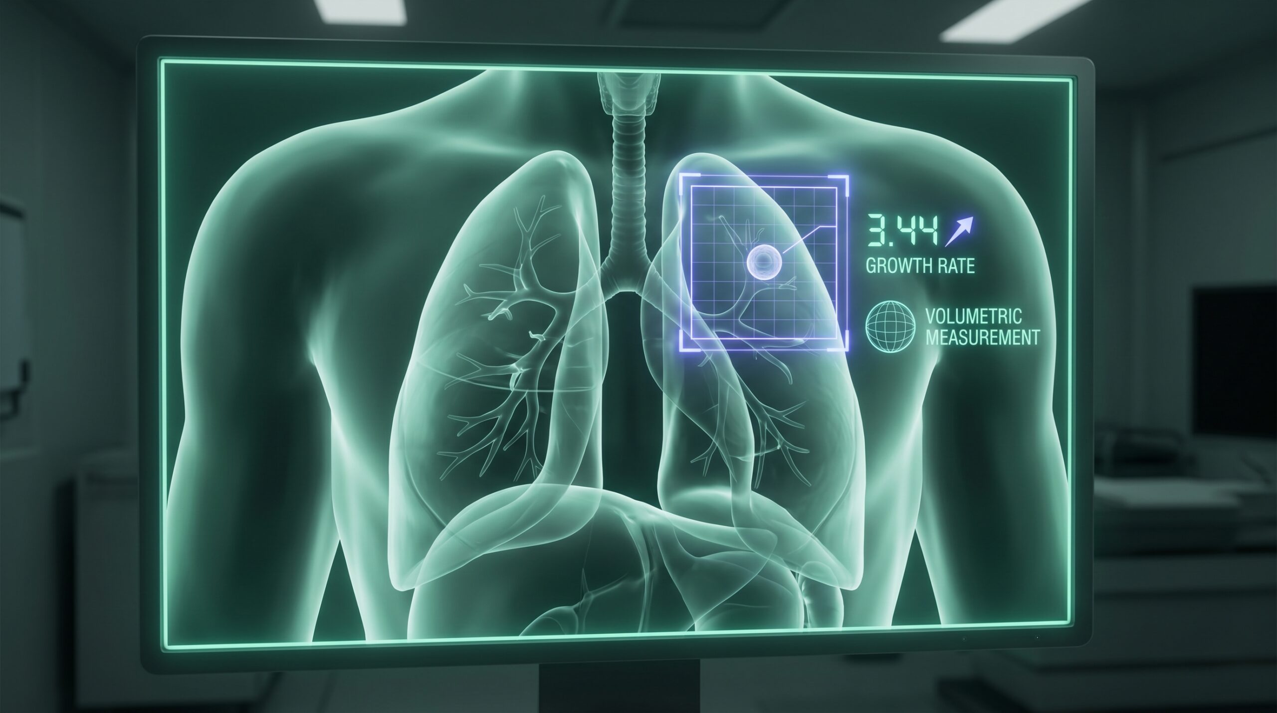
The American College of Radiology’s Lung Imaging Reporting and Data System (Lung-RADS) provides a structured framework for categorizing screening-detected nodules and guiding management — from category 1 (negative, no nodules) through category 4B (suspicious, ≥15mm solid nodules or growing nodules). AI tools increasingly integrate with Lung-RADS workflows, automatically measuring nodule dimensions, calculating growth rates on serial examinations, and suggesting Lung-RADS category assignments. This automated reporting reduces measurement variability between readers and ensures consistent protocol adherence across high-volume screening programs.
However, the limitations of diameter-based Lung-RADS criteria have become increasingly apparent. The NELSON trial’s volumetric approach — assessing nodule volume and volume doubling time (VDT) rather than diameter — produced substantially fewer false positives while maintaining sensitivity. The ESTI/ESR 2026 practice recommendations now explicitly state that deep learning algorithms are required for facilitating nodule detection and volumetric growth measurement in screening programs. This endorsement represents a significant milestone: for the first time, a major European guideline body has positioned AI not as optional but as a necessary component of screening infrastructure.
The shift from diameter to volume, and from static assessment to AI-powered temporal analysis, reflects a broader evolution toward risk-based screening management. AI algorithms that incorporate prior imaging data can track volumetric changes with greater precision and consistency than manual measurement, enabling more accurate VDT calculation and more confident management decisions. This is particularly valuable for nodules in the indeterminate range (Lung-RADS 3, 100–500 mm³ solid nodules) where the decision between short-term follow-up and invasive workup has significant implications for both patient outcomes and healthcare resource utilization.
Beyond Nodule Detection: Opportunistic Screening with AI
One of the most transformative applications of AI in lung cancer screening extends beyond the lungs themselves. LDCT scans of the thorax contain a wealth of anatomic information — including the heart, thoracic aorta, vertebral bodies, and body composition — that is typically underutilized in standard screening reads. AI algorithms can automatically extract these “opportunistic” biomarkers, providing clinically valuable information at no additional radiation exposure or cost to the patient.
Coronary artery calcification (CAC), the most common significant incidental finding on LDCT screening (reported in 12% of NLST participants), is a powerful predictor of cardiovascular events. Deep learning models trained on NLST data have demonstrated the ability to quantify CAC from non-gated LDCT with accuracy approaching dedicated cardiac CT. Given that cardiovascular disease was the leading cause of death in the NLST LDCT arm (26.1%, exceeding lung cancer deaths at 22.9%), automated CVD risk assessment from screening CT represents a compelling value proposition — particularly for the high-risk smoking population that shares cardiovascular and pulmonary risk factors.
Similarly, AI-based assessment of emphysema severity, vertebral compression fractures (indicating osteoporosis), and body composition (sarcopenia, visceral adiposity) from screening LDCT can identify patients who benefit from targeted interventions. This multiparametric approach aligns with the concept of the LDCT screening examination as a comprehensive health assessment rather than a single-disease test — an evolution that could dramatically improve the cost-effectiveness of screening programs and strengthen the case for broader implementation. The integration of AI into clinical workflows enables this expansion without increasing radiologist workload.
Expanding Access: Incidental Nodule Programs and AI
A particularly innovative approach to bridging the screening gap has emerged through incidental pulmonary nodule (IPN) programs — initiatives that leverage the rapidly growing volume of clinically indicated chest CTs (now 28 per 100 emergency department patients, up from 18 per 100 in 2006) to capture at-risk individuals outside of formal screening. Unlike traditional LDCT screening, which requires patients to meet specific eligibility criteria and actively enroll, IPN programs detect nodules on any chest CT performed for any indication, creating a much wider net for early lung cancer detection.
When coupled with AI-powered nodule detection and tracking systems, IPN programs have shown remarkable results. One US program reported diagnosing 7 to 10 times more lung cancer patients compared to traditional screening methods alone, capturing individuals who would not have met USPSTF eligibility criteria. AI plays a critical enabling role in these programs by automatically flagging nodules on all chest CTs, generating structured reports with Lung-RADS-compatible categories, and triggering follow-up management pathways — tasks that would be impractical to implement manually at scale.
The FDA has continued to clear new AI tools for this space, including the F.A.S.T. aiCockpit CT Lung Nodule system in January 2026, which integrates automated detection with workflow management and structured reporting. As the installed base of AI tools grows, the vision of universal lung nodule surveillance — rather than selective screening of a narrow eligible population — becomes increasingly feasible, potentially addressing the fundamental limitation that current guidelines capture only a fraction of those at risk.
Low-dose protocol (≤1 mSv) with deep learning reconstruction. Images auto-forwarded to AI platform and PACS simultaneously.
Deep learning algorithm identifies, segments, and measures all pulmonary nodules. Automated volumetry, morphology scoring, and Lung-RADS category assignment.
AI compares with prior imaging (if available) for VDT calculation. Malignancy probability score generated. Opportunistic biomarkers (CAC, emphysema) extracted.
Radiologist reviews AI output and assigns final Lung-RADS. Low-risk: annual follow-up. Intermediate: 3–6 month CT. High-risk: PET/CT or biopsy referral.
Radiation Dose Optimization and Deep Learning Reconstruction
The tension between minimizing radiation exposure and maintaining diagnostic image quality is a longstanding challenge in LDCT screening. Current guidelines recommend effective doses below 1 mSv — substantially less than a standard diagnostic chest CT (~7 mSv) but still non-negligible when applied to millions of individuals over decades of annual screening. Deep learning–based image reconstruction (DLR) has emerged as a critical enabling technology, allowing further dose reduction while preserving — or even enhancing — image quality compared to conventional filtered back projection (FBP) or iterative reconstruction methods.
A 2025 phantom study evaluating AI-based nodule detection across multiple reconstruction methods and ultra-low-dose protocols found that DLR maintained satisfactory detection sensitivity at significantly lower radiation doses than FBP or hybrid iterative reconstruction. At standard low-dose protocols (80 kV/160 mA), AI sensitivity exceeded 78% across all reconstruction methods, but at ultra-low-dose settings (80 kV/10 mA), only DLR maintained acceptable performance. These findings have direct implications for screening program design: the combination of deep learning reconstruction with AI-powered detection may enable “ultra-low-dose” screening protocols that further reduce the radiation burden on screened populations.
However, reconstruction parameters significantly influence AI performance. A 2025 study assessing AI detection of risk-dominant nodules in 300 LDCT scans from the NELCIN-B3 trial found that slice thickness, reconstruction kernel, and interval all affected detection accuracy. This dependency on technical parameters underscores the importance of standardizing acquisition and reconstruction protocols within screening programs — and of validating AI tools on the specific imaging protocols used at each site, rather than assuming that performance reported in one technical environment will transfer to another.
Future Directions
The next phase of AI-enhanced lung cancer screening is moving beyond detection and toward comprehensive risk orchestration. The NELSON-POP (Personalized Outcome Prediction) project exemplifies this direction, integrating genetic data (polygenic risk scores), environmental exposure (air pollution), AI-derived imaging biomarkers (nodule malignancy scores, emphysema quantification, coronary calcification), and individual clinical characteristics into multi-source prediction models. The goal is to stratify screening participants into personalized risk tiers, enabling individualized screening intervals, targeted management of detected nodules, and identification of high-risk individuals who may benefit from enhanced surveillance or chemoprevention.
Biomarker integration represents a complementary frontier. Models that combine circulating proteins or cell-free DNA with imaging features may achieve the precision needed to resolve the persistent false positive problem — particularly for sub-centimeter nodules where imaging alone has limited discriminatory power. The WCLC 2025 discussions signaled a fundamental shift from one-size-fits-all screening to personalized risk assessment combining biomarkers, AI analysis, genetic testing, and environmental factors.
For AI tools to fulfill their potential, regulatory and reimbursement frameworks must evolve. The FDA has cleared numerous AI diagnostic devices for radiology, with 295 new authorizations in 2025 alone. However, clinical validation in screening settings remains limited — most AI tools are validated on retrospective datasets rather than prospective screening cohorts. The LUNA25 Challenge, benchmarking AI against radiologists for lung nodule malignancy risk estimation on screening CT, represents an important step toward standardized, transparent evaluation of these tools. Until prospective, randomized evidence demonstrates that AI-enhanced screening improves lung cancer outcomes beyond what guideline-based LDCT alone achieves, the case for AI will rest on workflow efficiency and diagnostic accuracy rather than mortality reduction.
Clinical Implications
The evidence for AI-enhanced lung cancer screening is mature for nodule detection, promising for malignancy risk stratification, and early-stage for mortality impact. Radiologists and screening program directors should consider AI as an essential infrastructure component — not optional enhancement — given the ESTI/ESR 2026 endorsement and the mounting evidence that AI improves detection sensitivity, reduces measurement variability, and enables scalable screening operations. The choice of AI utilization model (assistant, prescreener, or backup) should be guided by each program’s priorities: maximizing cancer detection, minimizing false positives, or optimizing workflow efficiency.
For clinical practice, several considerations are paramount. First, AI tools for LDCT screening should be validated on the specific imaging protocols used at each site, given the demonstrated sensitivity of AI to reconstruction parameters. Second, the integration of malignancy risk prediction models — rather than simple detection — represents the highest-value AI application, as it directly addresses the false positive burden that undermines screening adherence and cost-effectiveness. Third, the expansion of AI capabilities beyond nodule detection to opportunistic cardiovascular and metabolic risk assessment transforms the LDCT screening examination into a comprehensive health evaluation, strengthening the clinical and economic rationale for broader implementation. Finally, clinicians should recognize that the 3–10% screening participation rate represents a far greater threat to population-level outcomes than any limitation of current AI technology — and that AI-enabled simplification of screening workflows, including automated eligibility assessment and patient outreach, may ultimately have greater impact than diagnostic refinement alone.
References
- National Lung Screening Trial Research Team. Reduced lung-cancer mortality with low-dose computed tomographic screening. N Engl J Med. 2011;365(5):395-409.
- de Koning HJ, van der Aalst CM, de Jong PA, et al. Reduced lung-cancer mortality with volume CT screening in a randomized trial (NELSON). N Engl J Med. 2020;382(6):503-513.
- Pinsky PF, Church TR, Izmirlian G, Kramer BS. Lung cancer incidence and mortality with extended follow-up in the National Lung Screening Trial. J Thorac Oncol. 2019;14(10):1732-1742.
- US Preventive Services Task Force. Screening for lung cancer: US Preventive Services Task Force recommendation statement. JAMA. 2021;325(10):962-970.
- Revel MP, Biederer J, Nair A, et al. ESR Essentials: lung cancer screening with low-dose CT — practice recommendations by the European Society of Thoracic Imaging. Eur Radiol. 2026;36:2064-2073. doi:10.1007/s00330-025-11910-9
- Khawaja A, Bartholmai BJ, Rajagopalan S, et al. A systematic review of AI performance in lung cancer detection on CT thorax. J Clin Med. 2025;14(13):4824.
- Kim Y, Yoon SH, Oh DY, et al. Artificial intelligence for low-dose CT lung cancer screening: comparison of utilization scenarios. AJR Am J Roentgenol. 2025;225(1). doi:10.2214/AJR.25.32829
- Venkadesh KV, Setio AAA, Schreuder A, et al. Prior CT improves deep learning for malignancy risk estimation of screening-detected pulmonary nodules. Radiology. 2023;308(1):e223308.
- Ardila D, Kiraly AP, Bharadwaj S, et al. End-to-end lung cancer screening with three-dimensional deep learning on low-dose chest computed tomography. Nat Med. 2019;25(6):954-961.
- Mikhael PG, Wohlwend J, Yala A, et al. Sybil: a validated deep learning model to predict future lung cancer risk from a single low-dose chest computed tomography. J Clin Oncol. 2023;41(12):2191-2200.
- Mao Y, Heuvelmans MA, van Tuinen M, et al. Artificial intelligence as an independent reader of risk-dominant lung nodules: influence of CT reconstruction parameters. Eur Radiol. 2025. doi:10.1007/s00330-025-11949-8
- Tanaka Y, Kubo T, Ohno Y, et al. Application of a pulmonary nodule detection program using AI technology to ultra-low-dose CT: differences in detection ability among various image reconstruction methods. Jpn J Radiol. 2025. doi:10.1007/s11604-025-01781-x
- Hassan OA, Podlaha T, Magee D, et al. Incidental pulmonary nodule programs working together with lung cancer screening and artificial intelligence to increase lung cancer detection. Cancers. 2025;17(4):685.
- Heuvelmans MA, Vonder M, Rook M, et al. Multi-source data approach for personalized outcome prediction in lung cancer screening: update from the NELSON trial (NELSON-POP). Eur J Epidemiol. 2023;38:575-581.
- Lu MT, Ivanov A, Mayrhofer T, et al. Deep learning to assess long-term mortality from chest radiographs. JAMA Netw Open. 2019;2(7):e197416.
- Zeleznik R, Foldyna B, Eslami P, et al. Deep learning predicts cardiovascular disease risks from lung cancer screening low dose computed tomography. Nat Commun. 2021;12:2963.
- F.A.S.T. aiCockpit CT Lung Nodule. FDA 510(k) Clearance. January 2026. K-number pending publication.
- Tushar FI, Pirmoazen AM, Dinnessen R, et al. Reproducible benchmarking for lung nodule detection and malignancy classification across multiple low-dose CT datasets. arXiv:2405.04605v5. February 2026.
Disclaimer: This article is intended for educational purposes and does not constitute medical advice. Lung cancer screening decisions should be individualized based on patient risk factors, preferences, and current institutional guidelines. MedTrainHub has no financial relationship with any AI platform manufacturer or screening guideline body mentioned in this article.
Conflicts of Interest: None declared.
Suggested Citation: MedTrainHub Editorial Team. Low-Dose CT Lung Cancer Screening: AI-Enhanced Nodule Detection and Risk Stratification. MedTrainHub.com. April 2026.